Theme images by kelvinjay. Powered by Blogger.

USA

AFRICA

ASIA

Brazil

Portugal

United Kingdom

Switzerland

New Cretaceous mammal fossil sheds light on evolution of middle ear


Researchers from the Institute of Vertebrate Paleontology and Paleoanthropology (IVPP) of the Chinese Academy of Sciences and the American Museum of Natural History (AMNH) have reported a new species of multituberculate - a type of extinct Mesozoic rodent - with well-preserved middle ear bones from the Cretaceous Jehol Biota of China. The findings were published in Nature.

New Cretaceous mammal fossil sheds light on evolution of middle ear
Reconstruction of Jeholbaatar kielanae [Credit: XU Yong]
The new mammal, Jeholbaatar kielanae, has a middle ear that is distinct from those of its relatives. WANG Yuanqing and WANG Haibing from IVPP, along with MENG Jin from AMNH, proposed that the evolution of its auditory apparatus might have been driven by specialization for feeding.

Fossil evidence shows that postdentary bones were either embedded in the postdentary trough on the medial side of the dentary or connected to the dentary via an ossified Meckel's cartilage in early mammals, prior to their migration into the cranium as seen in extant mammals.


Detachment of the mammalian middle ear bones from the dentary occurred independently at least three times. But how and why this process took place in different clades of mammals remains unclear.

The Jeholbaatar kielanae specimen was discovered in the Jiufotang Formation in China's Liaoning Province (Jehol Biota). It displays the first well-preserved middle-ear bones in multituberculates, providing solid evidence of the morphology and articulation of these bony elements, which are fully detached from the dentary.


It reveals a unique configuration with more complete components than those previously reported in multituberculates. The new fossil reveals a transitional stage in the evolution of the surangular - a "reptilian" jawbone.

In light of current evidence, scientists argue that the primary (malleus-incus) and secondary (squamosal-dentary) jaw joints co-evolved in allotherians, allowing a distinct palinal (anteroposterior) jaw movement while chewing.

Detachment of the auditory apparatus of the middle ear would have gained higher selective pressure in order to increase feeding efficiency, suggesting that evolution of the middle ear was probably triggered by functional constraints on the feeding apparatus in allotherians.

Source: Chinese Academy of Sciences [November 27, 2019]

New study shows a carnivorous dinosaur species regrew all its teeth every few months


A meat-eating dinosaur species that lived in Madagascar some 70 million years ago replaced all its teeth every couple of months or so, a new study has found, surprising even the researchers.

New study shows a carnivorous dinosaur species regrew all its teeth every few months
Credit: Sae Bom Ra
In fact, Majungasaurus grew new teeth roughly two to 13 times faster than those of other carnivorous dinosaurs, says paper lead author Michael D. D'Emic, an assistant professor of biology at Adelphi University. Majungasaurus would form a new tooth in each socket every couple of months.

"This meant they were wearing down their teeth quickly, possibly because they were gnawing on bones," D'Emic says. "There is independent evidence for this in the form of scratches and gouges that match the spacing and size of their teeth on a variety of bones - bones from animals that would have been their prey."


Some animals today, too, will gnaw on bones, including rodents, D'Emic explains. It's a way for them to ingest certain nutrients. It also requires exceptionally strong teeth - but Majungasaurus did not have those.

"That's our working hypothesis for why they had such elevated rates of replacement," D'Emic says. The rapid-fire tooth growth puts Majungasaurus in same league with sharks and big, herbivorous dinosaurs, he adds.

New study shows a carnivorous dinosaur species regrew all its teeth every few months
CT scan-generated models of the jaws of Majungasaurus (left), Ceratosaurus (center) and Allosaurus (right), with
microscopic views of the interior of their teeth below each model. Stripes running from upper left to lower right
in each microscopic image are daily deposited incremental lines, which allow the amount of time it took for
a tooth to grow to be reconstructed [Credit: D’Emic et al. 2019]
Although at least a few hundred meat-eating dinosaur species roamed the Earth, researchers have analyzed tooth-replacement rates for only about a half-dozen of them, D'Emic says. He also has looked into tooth-replacement patterns in plant-eating dinosaurs.

"I'm hoping this latest project spurs more people to study other species. I bet that will reveal further surprises," he says. "And hopefully that will lead to a better understanding of how dinosaurs evolved to be successful for so long."


Importantly, the recent study examined two additional species of predatory dinosaur (Allosaurus and Ceratosaurus), providing an opportunity to consider tooth-growth patterns at a broader scale.

In collaboration with Patrick O'Connor, professor of anatomy at Ohio University, and Ph.D. student Eric Lund, D'Emic used a collection of isolated fossil teeth to examine microscopic growth lines in the teeth. These growth lines are similar to tree rings, but instead of being deposited once a year, they were deposited daily. At the same, the team used computerized tomography (CT) on intact jaws to visualize unerupted teeth growing deep inside the bones. That allowed them to estimate tooth-replacement rates in a large number of individual jaws so they could cross-check their results.

People and climate led to Australia's megafauna extinction


The mystery of the role of people and climate in the fate of Australian megafauna might have been solved in a breakthrough study.

People and climate led to Australia's megafauna extinction
Pleistocene kangaroo was the largest and most heavily built kangaroo known
[Credit: Flinders University]
'Megafauna', giant beasts that once roamed the continent -- including wombat-like creatures as big as cars, birds more than two metres tall, and lizards more than seven metres long -- became extinct about 42,000 years ago. But the role of people in their demise has been hotly debated for decades.

For the first time, the research suggests a combination of climate change and the impact of people sealed the fate of megafauna, at least in south-eastern Australia. And that distribution of freshwater -- a precious commodity for animals and people alike as the climate warmed -- can explain regional differences in the timing at which megafauna died out.


The new study, led by a team of researchers from the ARC Centre of Excellence for Australian Biodiversity and Heritage (CABAH), analysed fossil data, climate reconstructions, and archaeological information describing patterns in human migration across south-eastern Australia.

The team developed and applied sophisticated mathematical models to test scenarios to explain regional variation in the periods during which people and megafauna coexisted.


"There has been much debate among scientists about what conditions led to this extinction event," said lead author Dr Frederik Saltre, Research Fellow and Coordinator of the Global Ecology Lab at Flinders University.

"Resolving this question is important because it is one of the oldest such extinction events anywhere after modern human beings evolved and left Africa", he added.


The findings, published in Nature Communications, are the result of analysis and complex modelling based on data including more than 10,000 fossils and archaeological records. Using high-quality fossil data and archaeological evidence of human activity, the researchers were able to map regional patterns of megafauna extinction.

They developed sophisticated models to test the impact of factors including climate, water availability, and human activity on localised patterns of megafauna extinction.

People and climate led to Australia's megafauna extinction
Credit: Flinders University
The extinction pattern could only be explained by the combination of people sharing the environment and the reduced of availability of freshwater due to climate change.

"The regional patterns in extinction are best explained by the hypothesis that people migrated across Australia, exploiting lakes and other sources of drinking water connecting the drier regions in between," said co-investigator Professor Corey Bradshaw of the Global Ecology Lab at Flinders University.

"It is plausible that megafauna species were attracted to the same freshwater sources as humans, thus increasing the chance of interactions."

The new insight that human pressure and climate change work together to trigger species extinction is a "stark warning" for the immediate future of the planet's biodiversity facing even stronger climate and habitat disruption, Dr Saltre concluded.

Source: Flinders University [November 27, 2019]

Animal-like embryos evolved before animals


Animals evolved from single-celled ancestors, before diversifying into 30 or 40 distinct anatomical designs. When and how animal ancestors made the transition from single-celled microbes to complex multicellular organisms has been the focus of intense debate.

Animal-like embryos evolved before animals
Three-dimensional reconstruction of a Caveasphaera specimen,
showing cell structures [Credit: NIGPAS]
Until now, this question could only be addressed by studying living animals and their relatives, but now the research team has found evidence that a key step in this major evolutionary transition occurred long before complex animals appear in the fossil record, in the fossilised embryos that resemble multicellular stages in the life cycle of single-celled relatives of animals.

The team discovered the fossils named Caveasphaera in 609 million-year old rocks in the Guizhou Province of South China. Individual Caveasphaera fossils are only about half a millimeter in diameter, but X-ray microscopy revealed that they were preserved all the way down to their component cells.


Kelly Vargas, from the University of Bristol's School of Earth Sciences, said: "X-Ray tomographic microscopy works like a medical CT scanner, but allows us to see features that are less than a thousandth of a millimeter in size. We were able to sort the fossils into growth stages, reconstructing the embryology of Caveasphaera."

Co-author Zongjun Yin, from Nanjing Institute of Geology and Palaeontology in China, added: "Our results show that Caveasphaera sorted its cells during embryo development, in just the same way as living animals, including humans, but we have no evidence that these embryos developed into more complex organisms."

Animal-like embryos evolved before animals
Computer models based on X-ray tomographic microscopy of the fossils, showing the successive stages
of development [Credit: Philip Donoghue & Zongjun Yin]
Co-author Dr John Cunningham, also from University of Bristol, said: "Caveasphaera had a life cycle like the close living relatives of animals, which alternate between single-celled and multicellular stages. However, Caveasphaera goes one step further, reorganising those cells during embryology."

Co-author Stefan Bengtson, from the Swedish Museum of Natural History, said "Caveasphaera is the earliest evidence of this most important step in the evolution of animals, which allowed them to develop distinct tissue layers and organs".


Co-author Maoyan Zhu, also from Nanjing Institute of Geology and Palaeontology, said he is not totally convinced that Caveasphaera is an animal. He added: "Caveasphaera looks a lot like the embryos of some starfish and corals - we don't find the adult stages simply because they are harder to fossilise

Co-author Dr Federica Marone from the Paul Scherrer Institute in Switzerland said "this study shows the amazing detail that can be preserved in the fossil record but also the power of X-ray microscopes in uncovering secrets preserved in stone without destroying the fossils."

a) and b), SEM image(s) of naked Caveasphaera specimen(s) with cellular structures;
e), f) showing detail of the cellular structures; c) and d), Caveasphaera specimens
with well-preserved envelope [Credit: NIGPAS]
Co-author Professor Philip Donoghue, also from the University of Bristol's School of Earth Sciences, said "Caveasphaera shows features that look both like microbial relatives of animals and early embryo stages of primitive animals. We're still searching for more fossils that may help us to decide.

"Either way, fossils of Caveasphaera tell us that animal-like embryonic development evolved long before the oldest definitive animals appear in the fossil record."

The findings are published in Current Biology.

Source: University of Bristol [November 27, 2019]

Researchers show how feathers propel birds through air and history


New research from an international team led by USC scientists set out to learn how feathers developed and helped birds spread across the world. Flight feathers, in particular, are masterpieces of propulsion and adaptation, helping penguins swim, eagles soar and hummingbirds hover.

Researchers show how feathers propel birds through air and history
A Taiwan blue magpie in flight [Credit: Shao Huan Lang]
Despite such diversity, the feather shares a common core design: a one-style-fits-all model with option trims for specialized performance. This simplicity and flexibility found in nature holds promise for engineers looking for better ways to build drones, wind turbines, medical implants and other advanced materials.

Those findings, published in Cell, offer an in-depth look at the form and function of a feather based on a comparative analysis of their physical structure, cellular composition and evolution. The study compares feathers of 21 bird species from around the world.

"We've always wondered how birds can fly in so many different ways, and we found the difference in flight styles is largely due to the characteristics of their flight feathers," said Cheng-Ming Chuong, the study's lead author and a developmental biologist in the Department of Pathology at the Keck School of Medicine of USC. "We want to learn how flight feathers are made so we can better understand nature and learn how biological architecture principles can benefit modern technology."


To gain a comprehensive understanding of the flight feather, Chuong formed a multi-disciplinary international team with Wen Tau Juan, a biophysicist at the Integrative Stem Cell Center, China Medical University in Taiwan. The work involved experts in stem cells, molecular biology, anatomy, physics, bio-imaging, engineering, materials science, bioinformatics and animal science. The bird species studied include ostrich, sparrow, eagle, chickens, ducks, swallow, owl, penguin, peacock, heron and hummingbird, among others.

They compared feathers using fossils, stem cells and flight performance characteristics. They focused on the feather shaft, or rachis, that supports the feather much like a mast holds a sail, bearing the stress between wind and wing. They also focused on the vane, the lateral branches astride the shaft that give the feather its shape to flap the air. And they examined how evolution shaped the barbs, ridges and hooks that help a feather hold its form and lock with adjacent feathers like Velcro to form a wing. The goal was to understand how a simple filament appendage on dinosaurs transformed into a three-level branched structure with different functions.

For birds such as ducks, eagles and sparrows that fly in different modes, the scientists noted significant differences in the feather shaft compared to ground-hugging birds. On the rigid exterior, the shaft cortex was thinner and lightweight, while the interior was filled with porous cells resembling bubble wrap, aligned into bands of various orientations and reinforced with ridges that operate like tiny lateral beams. Together, it forms a light, hollow and buoyant structure to enable flight. Cross-sections of feather shafts of different birds show highly specialized shapes and orientations of the inner core and outer cortex.

Researchers show how feathers propel birds through air and history
This picture shows a the asymmetric vane and tapering main shaft of a single flight feather
from a goshawk [Credit: Hao Howard Wu & Wen Tau Juan]
"The flight feather is made of two highly adaptable architectural modules, light and strong materials that can develop into highly adaptable configurations," Chuong said.

The researchers discovered two different molecular mechanisms guiding feather growth. Cortex thickness was governed by bone morphogenetic proteins, which are molecular signals for tissue growth. The porous feather interior, or medulla, relied upon a different mechanism known as transforming growth factor beta (TGF-b). Both components originate as stem cells in the bird's skin.

By contrast, feathers in flightless birds were simpler, consisting of a dense cortex exterior that is more rigid and sturdy with fewer internal struts and cells found in flying birds. The features were especially pronounced for penguins, which use wings as paddles under the water.


As part of the study, the researchers looked at nearly 100 million-year-old feathers, found embedded in amber in Myanmar. These fossils show early feathers lacked one key feature that modern birds have. Specifically, the researchers report how fossil feathers had barb branches and barbules, which form a feather vane by overlapping, but not hooklets. The hooklets, which act like clasps to turn fluffy feathers into a tight flat plane for high-performance flight, evolved later. The scientists also identified WNT2B, another growth factor, as the agent that controls hooklet formation. These also originated from epidermal stem cells.

Taken together, the findings show how feathered dinosaurs and early birds could form a primitive vane by overlapping barbule plates, although that wasn't aerodynamically fit to carry much load. As more complex composite features occurred in the wing, it got heavier, so feather shafts became stronger yet more lightweight, which led to stiffer feathers and sturdy wings that powered flight to carry birds around the world.

"Our findings suggest the evolutionary trends of feather shaft and vane are balanced for the best flight performance of an individual bird and become part of the selective basis of speciation," the study says. "The principles of functional architectures we studied here may also stimulate bio-inspired designs and fabrication of future composite materials for architectures of different scales, including wind turbines, artificial tissues, flying drones."

Source: University of Southern California [November 27, 2019]

Solving fossil mystery could aid quest for ancient life on Mars


Research which suggests that structures previously thought to be fossils may, in fact, be mineral deposits could save future Mars missions valuable time and resources.

Solving fossil mystery could aid quest for ancient life on Mars
A life-like structure created in the lab by chemical gardening
[Credit: Sean McMahon]
Microscopic tubes and filaments that resemble the remains of tiny creatures may have been formed by chemical reactions involving iron-rich minerals, the study shows. Previous research had suggested that such structures were among the oldest fossils on Earth.

The new findings could aid the search for extraterrestrial life during future missions to Mars by making it easier to distinguish between fossils and non-biological structures.

The discovery was made by a scientist from the University of Edinburgh who is developing techniques to seek evidence that life once existed on Mars.


Astrobiologist Sean McMahon created tiny formations in the lab that closely mimic the shape and chemical composition of iron-rich structures commonly found in Mars-like rocks on Earth, where some examples are thought to be around four billion years old.

Dr McMahon created the complex structures by mixing iron-rich particles with alkaline liquids containing the chemicals silicate or carbonate.

This process - known as chemical gardening - is thought to occur naturally where these chemicals abound. It can occur in hydrothermal vents on the seabed and when deep groundwater circulates through pores and fractures in rocks.


His findings suggest that structure alone is not sufficient to confirm whether or not microscopic life-like formations are fossils. More research will be needed to say exactly how they were formed.

The study, published in the journal Proceedings of the Royal Society B, was funded by the European Union's Horizon 2020 programme.

Dr Sean McMahon said: "Chemical reactions like these have been studied for hundreds of years but they had not previously been shown to mimic these tiny iron-rich structures inside rocks. These results call for a re-examination of many ancient real-world examples to see if they are more likely to be fossils or non-biological mineral deposits."

Source: University of Edinburgh [November 27, 2019]

World's oldest comma shrimp was way ahead of the curve


Scientists have discovered the world's oldest "comma" shrimp, a tiny crustacean shaped like its punctuation namesake. The 90-million-year-old creature fills in a major evolutionary gap for a family of marine animals now found in abundance around the planet, according to researchers from Yale and the University of Alaska. The discovery appears in the journal Proceedings of the Royal Society B.

World's oldest comma shrimp was way ahead of the curve
Eobodotria muisca [Credit: Javier Luque]
The fossilized shrimp, named Eobodotria muisca, comes from Mesozoic rocks in tropical South America. Researchers found exceptionally well-preserved fossils representing more than 500 individuals between 6 and 8 millimeters long, with features that are rarely preserved in fossil crustaceans: mouthparts, the gut, tiny hairs, and small compound eyes.

"We are amazed by how similar Eobodotria muisca is to today's species," said Yale paleontologist Javier Luque, lead author of the study. "There are eight families or main groups of living comma shrimp, and none of them have a confirmed fossil occurrence. This means we had no idea when modern comma shrimp evolved, until now."


The only previous record of a modern-looking comma shrimp is a 160-million-year-old fossil from Europe, Luque said. Although that shrimp fit within the range of comma shrimp body forms, it couldn't be linked to any of the main modern families of comma shrimp.

Eobodotria muisca, on the other hand, belongs to the Bodotriidae family of living comma shrimp, Luque said, extending the fossil record of that family of shrimp by nearly 100 million years.

The new species lived during the mid-Cretaceous period, when a long, narrow inland sea covered a large part of what is today the Eastern and Central Andes of Colombia. Luque found the fossils together with fossils of the crab Callichimaera perplexa. Eobodotria muisca is named after the Muisca native Americans who lived in the Colombian Andes.

Live 3-D model of Eobodotria muisca [Credit: Yale University]

Luque said that the similarity between Eobodotria muisca and its modern relatives suggests that the rates of external anatomical changes over millions of years in this group was low compared to other crustacean groups found in the same location. He also noted that the new cache of fossils is the first of its kind in northern South America.


Sarah Gerken of the University of Alaska-Anchorage is co-author of the study.

Gerken and Luque said most of the Eobodotria muisca fossils they found were adult males. Large aggregations of males usually happen in the water column when they are searching for females by means of their large antennae for smelling and their flappy tail appendages for swimming—both features that females lack, the researchers explained.

One possible explanation for this unusual accumulation of adult males is that they could have died suddenly in the water column while swarming in search of females, and then sunk down to the soft bottom where they fossilized, Gerken and Luque said.

The researchers said the discovery not only helps paleontologists understand the origin of the comma shrimp's curved body, it also can be used to help understand the origins of related crustaceans on the evolutionary family tree.

Author: Jim Shelton | Source: Yale University [November 26, 2019]

Did human hunting activities alone drive great auks' extinction?


New insight on the extinction history of a flightless seabird that vanished from the shores of the North Atlantic during the 19th century has been published in eLife.

Did human hunting activities alone drive great auks' extinction?
A mounted great auk skin, The Brussels Auk (RBINS 5355), from the collections
at the Royal Belgian Institute of Natural Sciences (RBINS)
[Credit: Thierry Hubin, RBINS]
The findings suggest that intense hunting by humans could have caused the rapid extinction of the great auk, showing how even species that exist in large and widespread populations can be vulnerable to exploitation.

Great auks were large, flightless diving birds thought to have existed in the millions. They were distributed around the North Atlantic, with breeding colonies along the east coast of North America and especially on the islands off Newfoundland. They could also be found on islands off the coasts of Iceland and Scotland, as well as throughout Scandinavia.


But these birds had a long history of being hunted by humans. They were poached for their meat and eggs during prehistoric times, and this activity was further intensified in 1500 AD by European seamen visiting the fishing grounds of Newfoundland. Their feathers later became highly sought after in the 1700s, contributing further to their demise.

"Despite the well-documented history of exploitation since the 16th century, it is unclear whether hunting alone could have been responsible for the species' extinction, or whether the birds were already in decline due to natural environmental changes," says lead author Jessica Thomas, who completed the work as part of her PhD studies at Bangor University, UK, and the University of Copenhagen, Denmark, and is now a postdoctoral researcher at Swansea University, Wales, UK.

Did human hunting activities alone drive great auks' extinction?
Great auk humeri from Funk Island. These samples are part of the great auk collection
at the American Museum of Natural History [Credit: J. Thomas]
To investigate this further, Thomas and her collaborators carried out combined analyses of ancient genetic data, GPS-based ocean current data, and population viability - a process that looks at the probability of a population going extinct within a given number of years. They sequenced complete mitochondrial genomes of 41 individuals from across the species' geographic range and used their analyses to reconstruct the birds' population structure and dynamics throughout the Holocene period, the last 11,700 years of Earth's history.

"Taken together, our data don't suggest that great auks were at risk of extinction prior to intensive human hunting behaviour in the early 16th century," explains co-senior author Thomas Gilbert, Professor of Evolutionary Genomics at the University of Copenhagen. "But critically, this doesn't mean that we've provided solid evidence that humans alone were the cause of great auk extinction. What we have demonstrated is that human hunting pressure was likely to have caused extinction even if the birds weren't already under threat from environmental changes."


Gilbert adds that their conclusions are limited by a couple of factors. The mitochondrial genome represents only a single genetic marker and, due to limited sample preservation and availability, the study sample size of 41 is relatively small for population genetic analyses.

"Despite these limitations, the findings help reveal how industrial-scale commercial exploitation of natural resources have the potential to drive an abundant, wide-ranging and genetically diverse species to extinction within a short period of time," says collaborator Gary Carvalho, Professor in Zoology (Molecular Ecology) at Bangor University. This echoes the conclusions of a previous study* on the passenger pigeon, a bird that existed in significant numbers before going extinct in the early 20th century.

"Our work also emphasises the need to thoroughly monitor commercially harvested species, particularly in poorly researched environments such as our oceans," concludes co-senior author Michael Knapp, Senior Lecturer in Biological Anthropology and Rutherford Discovery Fellow at the University of Otago, New Zealand. "This will help lay the platform for sustainable ecosystems and ensure more effective conservation efforts."

Source: eLife [November 26, 2019]

Dinosaur skull turns paleontology assumptions on their head


A team of researchers at the University of Alberta has unearthed a well-preserved Styracosaurus skull--and its facial imperfections have implications for how paleontologists identify new species of dinosaurs.

Dinosaur skull turns paleontology assumptions on their head
Styracosaurus skull [Credit: Pixabay]
The skull was discovered by Scott Persons in 2015, then a graduate student in the Department of Biological Sciences, during an expedition in the badlands northwest of Dinosaur Provincial Park.


Nicknamed Hannah, the dinosaur was a Styracosaurus - a horned dinosaur over five metres in length with a fan of long horns. UAlberta paleontologists led by Robert Holmes, professor in the Department of Biological Sciences, have learned much from those horns--because they aren't symmetrical.

"When parts of one side of the skull were missing, paleontologists have assumed that the missing side was symmetrical to the one that was preserved," explained Persons. "Turns out, it isn't necessarily. Today, deer often have left and right antlers that are different in terms of their branching patterns. Hannah shows dramatically that dinosaurs could be the same way."

Dinosaur skull turns paleontology assumptions on their head
Hannah's skull, seen from all sides. The jacket used to recover Hannah’s skull weighed 2500
kilograms—requiring a helicopter to retrieve from the field! [Credit: Scott Persons]
The differences in the skull's left and right halves are so extreme that had the paleontologists found only isolated halves, they might have concluded that they belong to two different species


"The skull shows how much morphological variability there was in the genus," said Holmes. Like the antlers of modern deer and moose, Hannah shows that the pattern of dinosaur horns could vary significantly--meaning some fossils that were once assumed to be unique species will have to be reevaluated.

Tradition dictates that the person who finds an important dinosaur specimen gets to give it a nickname. "Hannah the dinosaur is named after my dog," explained Persons, now a professor and museum curator at the College of Charleston. "She's a good dog, and I knew she was home missing me while I was away on the expedition."

Dinosaur skull turns paleontology assumptions on their head
Paleontologist Scott Persons, pictured alongside the partially-uncovered skull. The Styracosaurus skull
has implications for how horned dinosaurs are identified [Credit: Scott Persons]


Despite the nickname, paleontologists have no way of knowing if the dinosaur was female. But they have learned other details from the skull--from a partnership with researchers in the Faculty of Engineering.

"Ahmed Qureshi and graduate student Baltej Rupal in the Faculty of Engineering assisted us in performing a 3D laser scan of the skull," said Persons. "That let our publication to include a digital reconstruction, allowing scientists all over the world to download the 3D model and inspect it in detail."

"This is the future of paleontological collections: digital dinosaurs."

The paper was published in Cretaceous Research.

Author: Andrew Lyle | Source: University of Alberta [November 25, 2019]

16-million-year-old fossil shows springtails hitchhiking on winged termite


When trying to better the odds for survival, a major dilemma that many animals face is dispersal -- being able to pick up and leave to occupy new lands, find fresh resources and mates, and avoid intraspecies competition in times of overpopulation.

16-million-year-old fossil shows springtails hitchhiking on winged termite
Distribution of springtails on termite and ant hosts within ~ 16 Ma old Dominican amber
[Credit: N. Robin, C. D'Haese and P. Barden]
For birds, butterflies and other winged creatures, covering long distances may be as easy as the breeze they travel on. But for soil-dwellers of the crawling variety, the hurdle remains: How do they reach new, far-off habitats?

For one group of tiny arthropods called springtails (Collembola), a recent fossil discovery now suggests their answer to this question has been to piggyback on the dispersal abilities of others, literally.


In findings published in BMC Evolutionary Biology, researchers at the New Jersey Institute of Technology (NJIT) and Museum national d'Histoire naturelle have detailed the discovery of an ancient interaction preserved in 16-million-year-old amber from the Dominican Republic: 25 springtails attached to, and nearby, a large winged termite and ant from the days of the early Miocene.

The fossil exhibits a number of springtails still attached to the wings and legs of their hosts, while others are preserved as if gradually floating away from their hosts within the amber. Researchers say the discovery highlights the existence of a new type of hitchhiking behavior among wingless soil-dwelling arthropods, and could be key to explaining how symphypleonan springtails successfully achieved dispersal worldwide.

16-million-year-old fossil shows springtails hitchhiking on winged termite
Distribution of springtails on termite and ant hosts within ~ 16 Ma old Dominican amber, and illustration
of location of springtails on social insects [Credit: N. Robin, C. D’Haese and P. Barden]
"The existence of this hitchhiking behavior is especially exciting given the fact that modern springtails are rarely described as having any interspecfic association with surrounding animals," said Ninon Robin, the paper's first author whose postdoctoral research at NJIT's Department of Biological Sciences was funded by the Fulbright Program of the French-American Commission. "This finding underscores how important fossils are for telling us about unsuspected ancient ecologies as well as still ongoing behaviors that were so far simply overlooked."

Today, springtails are among the most common arthropods found in moist habitats around the world. Most springtails possess a specialized appendage under their abdomen they use to "spring" away in flee-like fashion to avoid predation. However this organ is not sufficient for traversing long distances, especially since most springtails are unable to survive long in dry areas.


The hitchhikers the researchers identified belong to a lineage of springtails found today on every continent, known as Symphypleona,which they say may have been "pre-adapted" to grasping on to other arthropods through prehensile antennae.

Because springtails would have encountered such winged termites and ants frequently due to their high abundance during the time of the preservation, these social insects may have been their preferred hosts for transportation.

16-million-year-old fossil shows springtails hitchhiking on winged termite
Allacma fusca is a species belonging to one of the three main groups of springtails today
known as Symphypleona [Credit: Urmas Tartes/Wikipedia]


"Symphypleonan springtails are unusual compared to other Collembola in that they have specialized antennae that are used in mating courtship," said Phillip Barden, assistant professor of biology at NJIT and the study's principal investigator. "This antennal anatomy may have provided an evolutionary pathway for grasping onto other arthropods. In this particular fossil, we see these specialized antennae wrapping around the wings and legs of both an ant and termite. Some winged ants and termites are known to travel significant distances, which would greatly aid in dispersal."

Barden says that the discovery joins other reports from the Caribbean and Europe of fossil springtails attached to a beetle, a mayfly and a harvestman in amber, which together suggest that this behavior may still exist today.

Barden notes that evidence of springtail hitchhiking may not have been captured in such high numbers until now due to the rarity of such a fossilized interaction, as well as the nature of modern sampling methods for insects, which typically involves submersion in ethanol for preservation.

"Because it appears that springtails reflexively detach from their hosts when in danger, evidenced by the detached individuals in the amber, ethanol would effectively erase the link between hitchhiker and host," said Barden. "Amber derives from fossilized sticky tree resin and is viscous enough that it would retain the interaction. ... Meaning, sometimes you have to turn to 16-million-year-old amber fossils to find out what might be happening in your backyard."

Author: Jesse Jenkins | Source: New Jersey Institute of Technology [November 25, 2019]

Aquatic microorganisms offer important window on the history of life


The air, earth and water of our planet are pulsating with living things. Yet, a vast and diverse web of life exists, about which almost nothing is known. This is the world of flagellates, tiny organisms that persist in staggering numbers in many diverse ecosystems around the world.

Aquatic microorganisms offer important window on the history of life
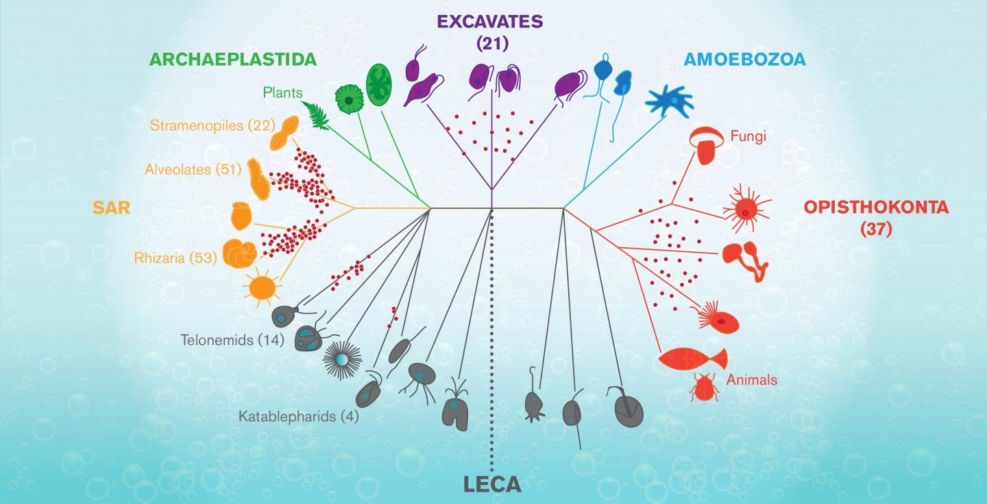
The graphic shows a tree of life for complex forms known as Eukaryotes, that arose mysteriously around 1.2-2 billions
years ago from a progenitor known as LECA (for Last Eukaryote Common Ancestor.) Jeremy Wideman and his
colleagues used a new method to sequence mitochondrial DNA for around 100 species of flagellates--tiny aquatic
organisms that populate many branches of the tree. These are seen on the graphic as red dots marking
the particular lineages these flagellates belong to [Credit: Shireen Dooling]
According to Jeremy Wideman, a researcher at the Biodesign Center for Mechanisms in Evolution at Arizona State University, we have a great deal to learn from these delicate and wildly varied creatures. Among other surprises, flagellates could provide valuable clues about a shadowy event that may have occurred 1.5-2 billion years ago, (no one is really sure of the timing), with the arrival of a new type of cell.

Known as LECA, it was a sort of primal egg out of which the astonishing profusion of complex life--from flagellate organisms, fungi and plants, to insects, zebra, and humans, exploded and spread over the earth.


In new research published in the journal Nature Microbiology, Wideman and his colleagues, including Prof. Thomas Richards at the University of Exeter describe a new method for investigating the genomes of eukaryotic flagellate organisms, which have been notoriously tricky to pinpoint and sequence.

Specifically, they explored samples of mitochondrial DNA, sequencing around 100 such genomes for previously undocumented flagellates. The new technique could help scientists like Wideman begin to fill in the largely blank region of the eukaryotic puzzle, where flagellate life flourishes.

Cellular worlds

Wideman, originally a traditional cell biologist, became frustrated with the many unaddressed questions in the field, recently joining the emerging discipline of evolutionary cell biology. This rapidly advancing research area uses cells as fundamental units for the study of evolutionary processes and imports concepts from evolutionary biology to better understand how cells work. "I'm literally a cell biologist that wants to know more about things we know nothing about," he says.

Evolutionary cell biology is a profoundly transdisciplinary endeavor, fusing evolutionary theory, genomics and cell biology with quantitative branches of biochemistry, biophysics, and population genetics.

Flagellates include many parasites implicated in human disease, from the intestinal bug Giardia to more damaging trypanosomes, and leishmania. Flagellates also perform more benevolent tasks. As the major consumers of bacteria and other protists in aquatic ecosystems, they help ensure the recycling of limiting nutrients.


Single-celled eukaryotic organisms, which include flagellates, constitute the overwhelming majority of eukaryotic diversity, vastly outpacing the more familiar multicellular plants, animals, and fungi. Despite their importance and ubiquity across the globe, flagellates are, as Wideman stresses, an almost entirely unknown inhabitant of the living world and one of the most enigmatic. When viewed under a microscope, their often science fiction-like appearance is markedly distinct from the kinds of eukaryotic cells commonly described in biology textbooks. Their emergence from comparatively rudimentary prokaryotes marks the most momentous transition in the history of life on earth.

"Novel lineages of heterotrophic flagellates are being discovered at an alarming, rate," Wideman says. "In the last two years 2 kingdom level lineages have been discovered (see here and here), meaning lineages that have been evolving independently of animals and fungi for over a billion years." Nevertheless, researchers have barely scratched the surface of this astonishing diversity and new methods must be brought to bear to speed up the quest. (Heterotrophs are organisms that cannot synthesize their own food, relying instead on other organisms for nutrition.)

Microbial safari

Any drop of pond, lake or ocean water is likely to contain many flagellates, but separating them from a multitude of non-flagellates and accurately reading their genomes by conventional means has been slow and painstaking work. Only a minute fraction of extant flagellates have known genomic sequences and it's even possible that the overwhelming majority have never actually been seen. According to Wideman, flagellate life forms represent the 'dark matter' of the eukaryotic universe.

"Heterotrophic flagellates are the target," Wideman says. "They're not a lineage. They're many, many lineages that are from all over the tree of life. LECA, the Last Eukaryotic Common Ancestor, was a heterotrophic flagellate, which means, that every major lineage (of eukaryotes) evolved from some sort of heterotrophic flagellate."


To access the elusive flagellate mitochondrial DNA, the researchers exploited a feature common to all flagellates and from which they take their name--the existence of flagella, which, unlike in animal sperm are on the front of cells and are often used to pull them forward like a microscopic breast stroke but are also involved in sensation, feeding, and perhaps other, as-yet unknown functions.

Flagella are rich in a particular protein known as tubulin. The new method for identifying flagellates and distinguishing them from their aquatic neighbors--primarily algae and bacteria--capitalizes on this fact by applying a selective stain to flagella-bearing organisms, activated by their high tubulin content. (Algal cells are naturally marked by their chloroplasts, which the flagellates of interest in the new study lack.)

Samples of sea water collected in 2014 off the coast of California provided a test case. Using the technique, the researchers gathered a windfall of mitochondrial sequence data, significantly expanding the catalog of flagellates identified by molecular means. Indeed, they doubled the existing mitochondrial DNA library for flagellate organisms. "We got many, many different kinds of organisms. So it was a very rich sample and very few were identical," Wideman says.

In search of LECA

Apart from the mystery of life's origin, the puzzle of where eukaryotes came from and how the LECA event transpired is the most important and vexing unanswered question in all of biology. (It has been dubbed the black hole at the heart of the living world.)

Correctly establishing the sequence of events underlying the crucial innovations within eukaryotes, from whence all complex life sprang, will take much more research in unexplored regions of the existing eukaryotic domain, particularly, the flagellates. Wideman believes the rapid advance of techniques for identifying and sequencing these organisms, such as the one outlined in the new study, offer hope such questions may one day find answers.

Author: Richard Harth | Source: Arizona State University [November 25, 2019]

New fossils shed light on how snakes got their bite and lost their legs


New fossils of an ancient legged snake, called Najash, shed light on the origin of the slithering reptiles.

New fossils shed light on how snakes got their bite and lost their legs
Lifelike rendition of Najash [Credit: Raul O. Gomez, Universidad de Buenos Aires]
The fossil discoveries published in Science Advances have revealed they possessed hind legs during the first 70 million years of their evolution. They also provide details about how the flexible skull of snakes evolved from their lizard ancestors.


The evolution of the snake body has captivated researchers for a long time - representing one of the most dramatic examples of the vertebrate body's ability to adapt - but a limited fossil record has obscured our understanding of their early evolution until now.

Dr Alessandro Palci, from Flinders University, was part of the international research team that performed high-resolution (CT) scanning and light microscopy of the preserved skulls of Najash to reveal substantial new anatomical data on the early evolution of snakes.

New fossils shed light on how snakes got their bite and lost their legs
Najash specimens from LBPA [Credit: Garberoglio et al. 2019]
"Snakes are famously legless, but then so are many lizards. What truly sets snakes apart is their highly mobile skull, which allows them to swallow large prey items. For a long time we have been lacking detailed information about the transition from the relatively rigid skull of a lizard to the super flexible skull of snakes".


"Najash has the most complete, three-dimensionally preserved skull of any ancient snake, and this is providing an amazing amount of new information on how the head of snakes evolved. It has some, but not all of the flexible joints found in the skull of modern snakes. Its middle ear is intermediate between that of lizards and living snakes, and unlike all living snakes it retains a well-developed cheekbone, which again is reminiscent of that of lizards."

Flinders University and South Australian Museum researcher Professor Mike Lee, was also part of the study, and adds "Najash shows how snakes evolved from lizards in incremental evolutionary steps, just like Darwin predicted."

New fossils shed light on how snakes got their bite and lost their legs
CT scan reconstructions of the articulated skull of Najash
[Credit: Garberoglio et al. 2019]
The new snake family tree also reveals that snakes possessed small but perfectly formed hind legs for the first 70 million years of their evolution.

"These primitive snakes with little legs weren't just a transient evolutionary stage on the way to something better. Rather, they had a highly successful body plan that persisted across many millions of years, and diversified into a range of terrestrial, burowing and aquatic niches," says Professor Lee.

Source: Flinders University [November 20, 2019]

Breakthrough method of identifying sex and species of million-year-old fossils


Reid Ferring, a professor in the University of North Texas Department of Geography and the Environment, is part of an international team of scientists who have developed a breakthrough method of identifying the sex and species of animal in fossils more than a million years old.

Breakthrough method of identifying sex and species of million-year-old fossils
UNT Professor Reid Ferring holds that cast of a skull found at the Dmanisi site
[Credit: University of North Texas]
"This is very exciting because our current method for determining sex and species, examination of extracted DNA, was limited to approximately 200,000 years. Through palaeoproteomics, the study of ancient proteins, we can now look back over a million years," said Ferring.

Ferring believes that palaeoproteomics will prove to be the key for establishing the evolutionary line between the earliest hominids and modern man. The reason, according to Ferring, is that proteins like collagen, which is found in tendons, ligaments, skin, bone and teeth, last much longer than DNA in fossilized material.


"We have thousands of hominid fossils in collections and museums around the world from all time periods," he said. "We have five complete skulls from the Dmanisi site in the country of Georgia that I know are almost two million years old. There are so many samples that can now be labeled and differentiated between species of the same line. We are on the edge of learning much more about our ancestors and ourselves than at any point in history."

Ferring added that the breakthrough came when the team was able to sample collagen from 1.7 million-year-old fossilized animal teeth found at the Dmanisi site. Using that protein, the team determined that the animal was a Stephanorhinus, an extinct form of rhinoceros. The team could then fit the Stephanorhinus into the modern rhinoceros' evolutionary line and differentiate it from ones that came before and after.


Palaeoproteomics is a very new field and, as such, scientists are very careful to document and confirm each part of the process. Ferring was one of more than 40 prominent researchers of different specialties, nationalities and backgrounds who participated in the groundbreaking project.

"I was brought in not because I am an expert on ancient proteins, but because I am a geologist and archaeologist who has been working at the Dmanisi site every summer for the last 27 years," Ferring said. "The whole surface of the site is covered with ruins of Bronze Age and medieval structures including a fortress and a seventh century Orthodox Church. All of the materials we excavated were found under 20 feet of volcanic ash containing thousands of animal bones and artifacts."


As the project geologist, Ferring documented and profiled the sediments in the area of the original find to provide context for all the materials that were dated and the fossils recovered. Based on the depth of the find and the type of minerals surrounding it, he was able to place the age of the Stephanorhinus teeth at more than 1.7 million years old.

In September, the journal Nature printed a paper titled "Early Pleistocene Enamel Proteome from Dmanisi Resolves Stephanorhinus Phylogeny" that describes the methods used and data collected by the international Stephanorhinus team. The paper was co-authored by Ferring.

Source: University of North Texas [November 19, 2019]

Entomologist claims photos show evidence of life on Mars


As scientists scramble to determine whether there is life on Mars, Ohio University Professor Emeritus William Romoser's research shows that we already have the evidence, courtesy of photographs from various Mars rovers.

Entomologist claims photos show evidence of life on Mars
A specimen whose head appears to have turned in the direction of the camera. Based on the scale provided in
the photo from which this was extracted, this individual is estimated to be approximately 20 inches long
 [Credit: Analysis by Dr. William Romoser]
Dr. Romoser, who specializes in arbovirology and general/medical entomology, has spent several years studying photographs from the red planet that are available on the Internet. He found numerous examples of insect-like forms, structured similarly to bees, as well as reptile-like forms, both as fossils and living creatures. He presented his findings at the national meeting of the Entomological Society of America in St. Louis, Missouri.


"There has been and still is life on Mars," Romoser said, noting that the images appear to show both fossilized and living creatures. "There is apparent diversity among the Martian insect-like fauna which display many features similar to Terran insects that are interpreted as advanced groups - for example, the presence of wings, wing flexion, agile gliding/flight, and variously structured leg elements."

Romoser said that while the Martian rovers, particularly the Curiosity Rover, have been looking for indicators of organic activity, there are a number of photos which clearly depict the insect- and reptile-like forms. Numerous photos show images where arthropod body segments, along with legs, antennae and wings, can be picked out from the surrounding area, and one even appears to show one of the insects in a steep dive before pulling up just before hitting the ground.

Entomologist claims photos show evidence of life on Mars
Putative fossil insect on its dorsum with head to the top, and with selected structures labelled
[Credit: Analysis by Dr. William Romoser]
Individual images were carefully studied while varying photographic parameters such as brightness, contrast, saturation, inversion, and so on. No content was added, or removed. Criteria used in Romoser's research included: Dramatic departure from the surroundings, clarity of form, body symmetry, segmentation of body parts, repeating form, skeletal remains, and observation of forms in close proximity to one another. Particular postures, evidence of motion, flight, apparent interaction as suggested by relative positions, and shiny eyes were taken to be consistent with the presence of living forms.


"Once a clear image of a given form was identified and described, it was useful in facilitating recognition of other less clear, but none-the-less valid, images of the same basic form," Romoser said. "An exoskeleton and jointed appendages are sufficient to establish identification as an arthropod. Three body regions, a single pair of antennae, and six legs are traditionally sufficient to establish identification as 'insect' on Earth. These characteristics should likewise be valid to identify an organism on Mars as insect-like. On these bases, arthropodan, insect-like forms can be seen in the Mars rover photos."

Entomologist claims photos show evidence of life on Mars
Ohio University Emeritus Professor William Romoser analyzed Mars rover photos and found
insect-like and reptile-like forms [Credit: Analysis by Dr. William Romoser]
Distinct flight behavior was evident in many images, Romoser said. These creatures loosely resemble bumble bees or carpenter bees on Earth. Other images show these "bees" appearing to shelter or nest in caves. And others show a fossilized creature that resembles a snake.


Romoser, who was an entomology professor at Ohio University for 45 years and co-founded its Tropical Disease Institute, also spent nearly 20 years as a visiting vector-borne disease researcher at the U.S. Army Medical Research Institute of Infectious Diseases. Between 1973 and 1998, Romoser authored and co-authored four editions of the widely-used textbook, "The Science of Entomology."

Romoser noted that interpretations of insect- and reptile-like creatures he described may change in the future as knowledge of life on Mars evolves, but that the sheer volume of evidence is compelling.

"The presence of higher metazoan organisms on Mars implies the presence of nutrient/energy sources and processes, food chains and webs, and water as elements functioning in a viable, if extreme, ecological setting sufficient to sustain life," he said. "I have observed instances suggestive of standing water or small water courses with evident meander and with the expected blurring of small submerged rocks, larger emergent rocks at the atmosphere/water interface, a moist bank area, and a drier area beyond the moist area. Water on Mars has been reported a number of times, including surface water detected by instrumentation on Viking, Pathfinder, Phoenix, and Curiosity.

"The evidence of life on Mars presented here provides a strong basis for many additional important biological as well as social and political questions," he added. It also represents a solid justification for further study."

Source: Ohio University [November 19, 2019]