Theme images by kelvinjay. Powered by Blogger.

USA

AFRICA

ASIA

Brazil

Portugal

United Kingdom

Switzerland

Researchers show how feathers propel birds through air and history


New research from an international team led by USC scientists set out to learn how feathers developed and helped birds spread across the world. Flight feathers, in particular, are masterpieces of propulsion and adaptation, helping penguins swim, eagles soar and hummingbirds hover.

Researchers show how feathers propel birds through air and history
A Taiwan blue magpie in flight [Credit: Shao Huan Lang]
Despite such diversity, the feather shares a common core design: a one-style-fits-all model with option trims for specialized performance. This simplicity and flexibility found in nature holds promise for engineers looking for better ways to build drones, wind turbines, medical implants and other advanced materials.

Those findings, published in Cell, offer an in-depth look at the form and function of a feather based on a comparative analysis of their physical structure, cellular composition and evolution. The study compares feathers of 21 bird species from around the world.

"We've always wondered how birds can fly in so many different ways, and we found the difference in flight styles is largely due to the characteristics of their flight feathers," said Cheng-Ming Chuong, the study's lead author and a developmental biologist in the Department of Pathology at the Keck School of Medicine of USC. "We want to learn how flight feathers are made so we can better understand nature and learn how biological architecture principles can benefit modern technology."


To gain a comprehensive understanding of the flight feather, Chuong formed a multi-disciplinary international team with Wen Tau Juan, a biophysicist at the Integrative Stem Cell Center, China Medical University in Taiwan. The work involved experts in stem cells, molecular biology, anatomy, physics, bio-imaging, engineering, materials science, bioinformatics and animal science. The bird species studied include ostrich, sparrow, eagle, chickens, ducks, swallow, owl, penguin, peacock, heron and hummingbird, among others.

They compared feathers using fossils, stem cells and flight performance characteristics. They focused on the feather shaft, or rachis, that supports the feather much like a mast holds a sail, bearing the stress between wind and wing. They also focused on the vane, the lateral branches astride the shaft that give the feather its shape to flap the air. And they examined how evolution shaped the barbs, ridges and hooks that help a feather hold its form and lock with adjacent feathers like Velcro to form a wing. The goal was to understand how a simple filament appendage on dinosaurs transformed into a three-level branched structure with different functions.

For birds such as ducks, eagles and sparrows that fly in different modes, the scientists noted significant differences in the feather shaft compared to ground-hugging birds. On the rigid exterior, the shaft cortex was thinner and lightweight, while the interior was filled with porous cells resembling bubble wrap, aligned into bands of various orientations and reinforced with ridges that operate like tiny lateral beams. Together, it forms a light, hollow and buoyant structure to enable flight. Cross-sections of feather shafts of different birds show highly specialized shapes and orientations of the inner core and outer cortex.

Researchers show how feathers propel birds through air and history
This picture shows a the asymmetric vane and tapering main shaft of a single flight feather
from a goshawk [Credit: Hao Howard Wu & Wen Tau Juan]
"The flight feather is made of two highly adaptable architectural modules, light and strong materials that can develop into highly adaptable configurations," Chuong said.

The researchers discovered two different molecular mechanisms guiding feather growth. Cortex thickness was governed by bone morphogenetic proteins, which are molecular signals for tissue growth. The porous feather interior, or medulla, relied upon a different mechanism known as transforming growth factor beta (TGF-b). Both components originate as stem cells in the bird's skin.

By contrast, feathers in flightless birds were simpler, consisting of a dense cortex exterior that is more rigid and sturdy with fewer internal struts and cells found in flying birds. The features were especially pronounced for penguins, which use wings as paddles under the water.


As part of the study, the researchers looked at nearly 100 million-year-old feathers, found embedded in amber in Myanmar. These fossils show early feathers lacked one key feature that modern birds have. Specifically, the researchers report how fossil feathers had barb branches and barbules, which form a feather vane by overlapping, but not hooklets. The hooklets, which act like clasps to turn fluffy feathers into a tight flat plane for high-performance flight, evolved later. The scientists also identified WNT2B, another growth factor, as the agent that controls hooklet formation. These also originated from epidermal stem cells.

Taken together, the findings show how feathered dinosaurs and early birds could form a primitive vane by overlapping barbule plates, although that wasn't aerodynamically fit to carry much load. As more complex composite features occurred in the wing, it got heavier, so feather shafts became stronger yet more lightweight, which led to stiffer feathers and sturdy wings that powered flight to carry birds around the world.

"Our findings suggest the evolutionary trends of feather shaft and vane are balanced for the best flight performance of an individual bird and become part of the selective basis of speciation," the study says. "The principles of functional architectures we studied here may also stimulate bio-inspired designs and fabrication of future composite materials for architectures of different scales, including wind turbines, artificial tissues, flying drones."

Source: University of Southern California [November 27, 2019]
Aquatic microorganisms offer important window on the history of life


The air, earth and water of our planet are pulsating with living things. Yet, a vast and diverse web of life exists, about which almost nothing is known. This is the world of flagellates, tiny organisms that persist in staggering numbers in many diverse ecosystems around the world.

Aquatic microorganisms offer important window on the history of life
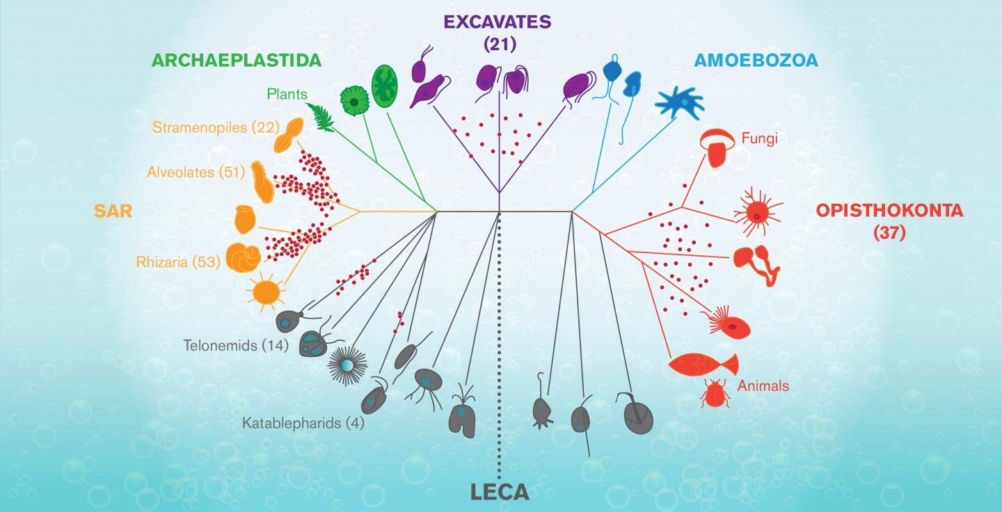
The graphic shows a tree of life for complex forms known as Eukaryotes, that arose mysteriously around 1.2-2 billions
years ago from a progenitor known as LECA (for Last Eukaryote Common Ancestor.) Jeremy Wideman and his
colleagues used a new method to sequence mitochondrial DNA for around 100 species of flagellates--tiny aquatic
organisms that populate many branches of the tree. These are seen on the graphic as red dots marking
the particular lineages these flagellates belong to [Credit: Shireen Dooling]
According to Jeremy Wideman, a researcher at the Biodesign Center for Mechanisms in Evolution at Arizona State University, we have a great deal to learn from these delicate and wildly varied creatures. Among other surprises, flagellates could provide valuable clues about a shadowy event that may have occurred 1.5-2 billion years ago, (no one is really sure of the timing), with the arrival of a new type of cell.

Known as LECA, it was a sort of primal egg out of which the astonishing profusion of complex life--from flagellate organisms, fungi and plants, to insects, zebra, and humans, exploded and spread over the earth.


In new research published in the journal Nature Microbiology, Wideman and his colleagues, including Prof. Thomas Richards at the University of Exeter describe a new method for investigating the genomes of eukaryotic flagellate organisms, which have been notoriously tricky to pinpoint and sequence.

Specifically, they explored samples of mitochondrial DNA, sequencing around 100 such genomes for previously undocumented flagellates. The new technique could help scientists like Wideman begin to fill in the largely blank region of the eukaryotic puzzle, where flagellate life flourishes.

Cellular worlds

Wideman, originally a traditional cell biologist, became frustrated with the many unaddressed questions in the field, recently joining the emerging discipline of evolutionary cell biology. This rapidly advancing research area uses cells as fundamental units for the study of evolutionary processes and imports concepts from evolutionary biology to better understand how cells work. "I'm literally a cell biologist that wants to know more about things we know nothing about," he says.

Evolutionary cell biology is a profoundly transdisciplinary endeavor, fusing evolutionary theory, genomics and cell biology with quantitative branches of biochemistry, biophysics, and population genetics.

Flagellates include many parasites implicated in human disease, from the intestinal bug Giardia to more damaging trypanosomes, and leishmania. Flagellates also perform more benevolent tasks. As the major consumers of bacteria and other protists in aquatic ecosystems, they help ensure the recycling of limiting nutrients.


Single-celled eukaryotic organisms, which include flagellates, constitute the overwhelming majority of eukaryotic diversity, vastly outpacing the more familiar multicellular plants, animals, and fungi. Despite their importance and ubiquity across the globe, flagellates are, as Wideman stresses, an almost entirely unknown inhabitant of the living world and one of the most enigmatic. When viewed under a microscope, their often science fiction-like appearance is markedly distinct from the kinds of eukaryotic cells commonly described in biology textbooks. Their emergence from comparatively rudimentary prokaryotes marks the most momentous transition in the history of life on earth.

"Novel lineages of heterotrophic flagellates are being discovered at an alarming, rate," Wideman says. "In the last two years 2 kingdom level lineages have been discovered (see here and here), meaning lineages that have been evolving independently of animals and fungi for over a billion years." Nevertheless, researchers have barely scratched the surface of this astonishing diversity and new methods must be brought to bear to speed up the quest. (Heterotrophs are organisms that cannot synthesize their own food, relying instead on other organisms for nutrition.)

Microbial safari

Any drop of pond, lake or ocean water is likely to contain many flagellates, but separating them from a multitude of non-flagellates and accurately reading their genomes by conventional means has been slow and painstaking work. Only a minute fraction of extant flagellates have known genomic sequences and it's even possible that the overwhelming majority have never actually been seen. According to Wideman, flagellate life forms represent the 'dark matter' of the eukaryotic universe.

"Heterotrophic flagellates are the target," Wideman says. "They're not a lineage. They're many, many lineages that are from all over the tree of life. LECA, the Last Eukaryotic Common Ancestor, was a heterotrophic flagellate, which means, that every major lineage (of eukaryotes) evolved from some sort of heterotrophic flagellate."


To access the elusive flagellate mitochondrial DNA, the researchers exploited a feature common to all flagellates and from which they take their name--the existence of flagella, which, unlike in animal sperm are on the front of cells and are often used to pull them forward like a microscopic breast stroke but are also involved in sensation, feeding, and perhaps other, as-yet unknown functions.

Flagella are rich in a particular protein known as tubulin. The new method for identifying flagellates and distinguishing them from their aquatic neighbors--primarily algae and bacteria--capitalizes on this fact by applying a selective stain to flagella-bearing organisms, activated by their high tubulin content. (Algal cells are naturally marked by their chloroplasts, which the flagellates of interest in the new study lack.)

Samples of sea water collected in 2014 off the coast of California provided a test case. Using the technique, the researchers gathered a windfall of mitochondrial sequence data, significantly expanding the catalog of flagellates identified by molecular means. Indeed, they doubled the existing mitochondrial DNA library for flagellate organisms. "We got many, many different kinds of organisms. So it was a very rich sample and very few were identical," Wideman says.

In search of LECA

Apart from the mystery of life's origin, the puzzle of where eukaryotes came from and how the LECA event transpired is the most important and vexing unanswered question in all of biology. (It has been dubbed the black hole at the heart of the living world.)

Correctly establishing the sequence of events underlying the crucial innovations within eukaryotes, from whence all complex life sprang, will take much more research in unexplored regions of the existing eukaryotic domain, particularly, the flagellates. Wideman believes the rapid advance of techniques for identifying and sequencing these organisms, such as the one outlined in the new study, offer hope such questions may one day find answers.

Author: Richard Harth | Source: Arizona State University [November 25, 2019]

Unravelling gene expression


The DNA of a single cell is 2-3 meters long end-to-end. To fit and function, DNA is packaged around specialized proteins. These DNA-protein complexes are called nucleosomes, and they are a small part of a larger structure called chromatin. Nucleosomes can be thought of as the cell's DNA storage and protection unit.

Unravelling gene expression
The pioneer transcription factor Rap1 pries open compact chromatin
structure to activate genes [Credit: Beat Fierz, EPFL]
When a particular gene needs to be expressed, the cell requires access to the protected DNA within chromatin. This means that the chromatin structure must be opened and the nucleosomes must be removed to expose the underlying target gene.


This takes place in the orchestrated process of "chromatin remodeling", which regulates gene expression and involves a multitude of actors. Unravelling this pivotal step not only furthers our fundamental understanding, but may also help in the development of genetic engineering tools.

Now the lab of Beat Fierz at EPFL, has been able to uncover the first steps in the chromatin-opening process at the level of a single molecule, using a combination of chemical biology and biophysical methods. Published in Molecular Cell, the work looks at the role of a group of proteins called "pioneer transcription factors". These proteins bind to specific DNA regions within chromatin that are themselves shielded from other proteins. Little is known about how these factors overcome the barriers of the chromatin maze.

Unravelling gene expression
The graphical abstract of the paper, showing the way Rap1 accesses chromatin
[Credit: M. Mivelaz et al. 2019]


Fierz's lab looked at yeast, which is a model organism for human genetics. The method involved replicating the architecture of yeast genes, combined with single-molecule fluorescence. The researchers studied a yeast pioneer transcription factor called Rap1, and found that it choreographs chromatin remodeling, allowing access to other proteins required for gene expression that were previously obstructed.

To do this, Rap1 first binds chromatin and then influences the action of a large molecular machine called "Remodeling the Structure of Chromatin" (RSC), displacing nucleosomes and paving the way to the now-exposed DNA for other proteins involved in controlling gene expression.

By revealing the physico-chemical mechanism of how Rap1 gains access to chromatin and opens it up, the EPFL study proposes a biological model for other pioneer transcription factors, but also provides the tools for investigating them at the level of a single molecule.

Source: Ecole Polytechnique Federale de Lausanne [November 21, 2019]

Self-restrained genes enable evolutionary novelty


Changes in the genes that control development can potentially make large contributions to evolution by generating new morphologies in plants and animals. However, because developmental genes frequently influence many different processes, changes to their expression carry a risk of "collateral damage". Scientists at the Max Planck Institute for Plant Breeding Research in Cologne, and collaborators, have now shown how gene self-repression can reduce the potential side effects of novel gene expression so that new forms can evolve. This self-regulation occurs via a distinctive molecular mechanism employing small regions of genomic DNA called low-affinity transcription factor binding sites.

Self-restrained genes enable evolutionary novelty
A confocal micrograph of a young leaf of Cardamine hirsuta (hairy bittercress) with
emerging leaflets, showing distribution of the RCO protein. Cell outlines are shown in gray.
RCO shown here in red colour is active at the base of initiating leaflets where it reduces
 growth, leading to the formation of leaflets that are separated from each other
 [Credit: Neha Bhatia and Peter Huijser]
Suppose a bird develops a modified wing shape, which makes flying easier and could be beneficial to its survival. If this gene change also altered the bird's color, making it less attractive to mates, then the advantageous wing-shape modification would be unlikely to persist. So, how then does nature balance the potential for novelty, with the risk of side effects that may prevent novelty from arising? Using the evolution of leaf shape as an example, an international team led by Director Miltos Tsiantis has provided fresh insight into this question.


This new study was done in the hairy bittercress, a small weed that the Tsiantis group has developed into a model system for understanding evolution of plant form. It builds on previous work from the group in which a gene called RCO was found to have driven leaf shape diversification in mustard plants by acquiring a novel expression pattern.

RCO encodes a transcription factor, a type of protein that can turn other genes on or off, and RCO's new expression pattern resulted in the emergence of the more complex leaf shapes found in bittercress. The researchers have now shown that this change in gene expression was accompanied by RCO acquiring the ability to repress its own activity. Mike Levine, Director of the Lewis-Sigler Institute for Integrative Genomics at Princeton University who was not involved in the study, finds this particular insight "very compelling". As the self-repression of RCO "limits the scope of its activity", Levine explains, it "thereby blocks potentially deleterious influences on cell development and function".

Stimulating cytokinin

As a next step, the scientists identified the genes targeted by RCO, and found that many of them are responsible for coordinating local levels of cytokinin - a widely acting plant hormone known to affect cell growth. Importantly, when the self-regulation of RCO is modified, RCO stimulates cytokinin excessively and leaf shape is altered in ways that can negatively affect plant fitness. This finding confirms the idea that self-repression of RCO could be essential for the persistence of RCO-induced novel leaf morphologies.


What's particularly interesting is that this self-repression of RCO occurs in a very distinctive way. The scientists discovered that it is based on many weak interactions between the RCO protein and RCO regulatory DNA at low-affinity binding sites. "This finding is exciting", explains Tsiantis, "because low-affinity binding sites can evolve relatively quickly, thus offering an easy way for evolution to keep changes in gene expression in check, by lowering a regulator's expression".

Soft repression

Indeed, this latest work from Tsiantis's team directly demonstrates that low-affinity transcription factor binding sites can play a major role in the generation of morphological novelty. By providing a tool to "softly" repress RCO expression, these sites dampen the effects of RCO expression changes and allow cytokinin levels to be fine-tuned. This in turn promotes the appearance of more complex leaf shapes, e.g., by precisely regulating the outgrowth of lobes or leaflets along the margins of developing leaves.

These results will stimulate further efforts to understand the influence of low-affinity transcription factor binding sites on development, diversity and disease. For example, there is increasing awareness that changes in the regulation of developmental genes are a major contributor to human disease, and that other regulatory changes can reduce disease severity or protect individuals who carry disease variants. While the specific DNA sequences underlying these effects are often unknown, this latest work highlights low-affinity transcription factor binding sites as excellent candidate regions for identifying causal sequences of disease susceptibility, and for understanding variation in trait diversity more broadly in complex eukaryotes.

The study is published in Current Biology.

Source: Max Planck Society [November 21, 2019]

Researchers sequence genome of the 'devil worm'


When scientists discovered a worm deep in an aquifer nearly one mile underground, they hailed it as the discovery of the deepest-living animal ever found. Now American University researchers, reporting in Nature Communications, have sequenced the genome of the unique animal, referred to as the 'Devil Worm' for its ability to survive in harsh, subsurface conditions. The Devil Worm's genome provides clues to how an organism adapts to lethal environmental conditions. Future research into how it evolved could help humans learn lessons for how to adapt to a warming climate.

Researchers sequence genome of the 'devil worm'
H. Mephisto (the Devil Worm) (microscopic image, magnified 200x)
[Credit: Prof. John Bracht, American University]
In 2008, Gaetan Borgonie from the University of Ghent and Princeton University geoscientist Tullis Onstott discovered the microscopic Devil Worm while investigating subterrestrial bacterial communities in active gold mines in South Africa. Borgonie and his team were stunned to discover the worm, a complex, multi-cellular animal thriving in an environment thought only livable for microbes, with high temperatures, little oxygen and high amounts of methane.

Researchers named the worm Halicephalobus mephisto, in honor of Mephistopheles, a subterranean demon from the medieval German legend Faust.

The Devil Worm is the first subterrestrial animal to have its genome sequenced. The genome offers evidence of how life can exist below Earth's surface and opens up a new way of understanding how life can survive beyond Earth, said John Bracht, assistant professor of biology at American University who led the genome sequencing project.


The sequencing revealed that the genome encodes an unusually large number of heat-shock proteins known as Hsp70, which is notable because many nematode species whose genomes are sequenced do not reveal such a large number. Hsp70 is a well-studied gene that exists in all life forms and restores cellular health due to heat damage.

Many of the Hsp70 genes in the Devil Worm's genome were copies of themselves. The genome also has extra copies of AIG1 genes, known cellular survival genes in plants and animals. More research will be needed, but Bracht believes the presence of copies of the gene signifies the worm's evolutionary adaptation.

"The Devil Worm can't run away; it's underground," Bracht explained. "It has no choice but to adapt or die. We propose that when an animal cannot escape intense heat, it starts making additional copies of these two genes to survive."


By scanning other genomes, Bracht identified other cases where the same two gene families, Hsp70 and AIG1, are expanded. The animals he identified are bivalves, a group of mollusks including clams, oysters and mussels. They are heat-adapted like the Devil Worm. This suggests that the pattern identified in the Devil Worm may extend more generally to organisms unable to escape environmental heat. This work was also recently published in the Journal of Molecular Evolution with an AU Biology undergraduate first author, Megan Guerin.

Bracht got the chance to sequence the unique worm's genome as a post-doctoral fellow at Princeton University. He carried the project over to American University when he joined the biology faculty in 2014. Two biology master's students working in his lab, Deborah Weinstein and Sarah Allen, contributed research and writing of the Nature Communications paper and are first and second authors, respectively, on the manuscript. Kathryn Walters-Conte, Ph.D., director of AU's Master's in Biotechnology program, also contributed to the paper.

Nearly a decade ago, the Devil Worm was unknown and living below the Earth's surface. Now it's a subject of study in science laboratories, including Bracht's. When Bracht brought Devil Worms from a laboratory in South Africa that cultures them to his laboratory at AU, he recalls saying to his students that aliens landed at AU. The metaphor isn't a stretch. NASA supports research of the Devil Worm for what it can teach scientists about the search for life beyond Earth.


"Part of this work entails looking for 'biosignatures' of life--stable chemical clues left behind by living things. We focus on a ubiquitous biosignature of organic life--genomic DNA--obtained from an animal that has adapted to an environment once considered uninhabitable to complex life: the deep terrestrial subsurface," Bracht said. "It is work that might prompt us to broaden the search for extraterrestrial life to 'uninhabitable' exoplanets' deep subterrestrial regions."

Nematodes are well suited to studies of evolutionary adaptation, Bracht said. They have adapted to a diverse set of environments and are among the most abundant animals on earth. Future work involving the Devil Worm in Bracht's lab will pinpoint Hsp70's function, such as inactivating the gene to test its response to heat stress. Other work could involve gene-transfer studies in C.elegans, a type of heat-intolerant microscopic roundworm, to see if it becomes heat-resilient.

Source: American University [November 21, 2019]

Coming to a Head: Insights from a Vampire of the Deep


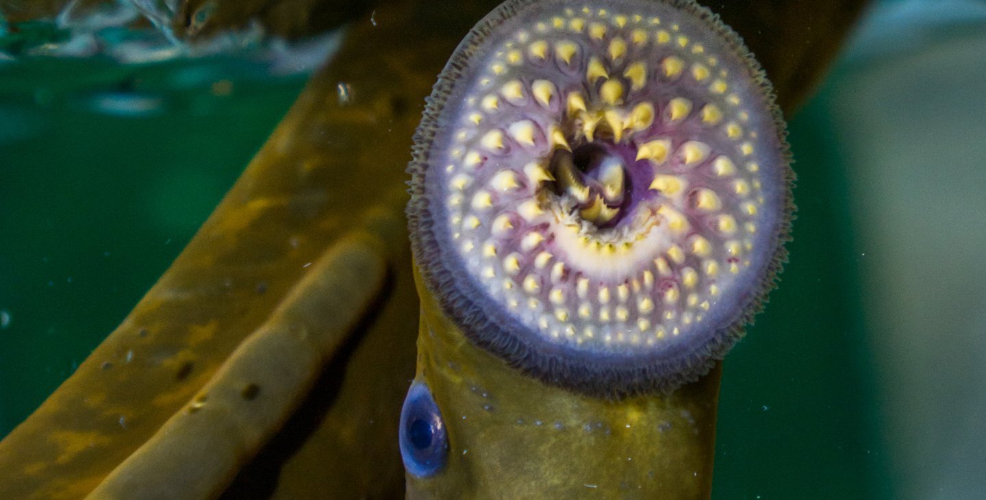
Lamprey are blood-sucking vampire-like fish that attach to and eventually kill game fish, making them the bane of many a fisherman's existence. Like something out of a horror film, these parasites use radial rows of sharp teeth to dig into the skin of their host and extract blood and other fluids for food. But to Caltech scientists, these gruesome pests hold important clues to the evolution and success of vertebrates.

Coming to a Head: Insights from a Vampire of the Deep
Credit: Megan Martik
The eel-like fish do not have a jaw, but because lamprey are the most primitive animals with a backbone (vertebrates) on Earth, comparing them to more recently evolved vertebrates could offer insight into how the jaw and other structures of the head evolved over time. This has important implications for understanding behaviors such as predation that rely heavily on the power of the jaw.


Caltech scientists have discovered that a population of embryonic stem cells called neural crest cells may help explain how vertebrates progressively evolved a more and more efficient head. Neural crest cells originate within the developing central nervous system before migrating throughout the embryo. They are a major player in the development of the jaws and other important components of the head and face, making them a good candidate for a role in vertebrate head evolution.

The research was conducted in the laboratory of Marianne Bronner, Albert Billings Ruddock Professor of Biology and director of the Beckman Institute. A paper describing work appeared in the journal Nature.

"Lamprey are like living fossils, and we use them to guess what primitive vertebrates might have looked like," says Bronner. "In the 1980s, it was proposed that the neural crest was a vertebrate invention that enabled acquisition of a 'new head.' We wanted to test this by looking at neural crest genes in lamprey."


The study, led by postdoctoral scholar Megan Martik, began by comparing the genetic makeup of lamprey neural crest with that of chicken embryos. The team discovered that the lamprey neural crest cells lacked important genes that are present in the head neural crest of birds and mammals, and critical for making advanced structures like the jaw.

The team wondered how the neural crest ultimately acquired the genes. To look for clues, they examined the neural crest genes of animals that have jaws, such as sharks and zebrafish, and evolved after lamprey but well before chickens and other birds. Martik and her colleagues found that the genetic program that helped to make the head more sophisticated was created by the progressive addition of genes to the neural crest repertoire as the vertebrate tree of life advanced and new species evolved.

Author: Alison Koontz | Source: California Institute of Technology [November 18, 2019]

New method takes analysis of genetic libraries to next level


Uppsala researchers have developed a new method for investigating dynamic processes in large genetic libraries. By using this method to study cell cycle regulation, they help paint a clearer picture of the elusive control mechanism. The study is published in the journal Nature Methods.

New method takes analysis of genetic libraries to next level
Credit: Bill Oxford/Getty Images
Modern gene technology makes it possible to quickly and inexpensively introduce thousands of different DNA modifications in human cells or bacteria to create genetic libraries. The CRISPR/Cas9 system, a.k.a. 'the gene snipper', can be modified and used to alter the expression of thousands of different proteins. By labelling each modification with a genetic barcode, it is possible to keep track of which cell carries which change.

At the same time, recent developments in optics and image analysis have made it possible to investigate the chemical processes inside the cell with exceedingly high precision. In principle, it is possible to 'film' basic biological processes such as protein expression or cell division at the molecular level inside a living cell.


Now imagine it were possible to combine these advanced optical methods with large-scale genetic engineering. Let us say we are interested in a particular biological process. We could, in theory, identify all the genes involved in this process by observing the biology in a genetic library. Studies that have so far taken several years could be conducted in a single experiment - in theory.

The challenges that have previously prevented scientists from putting theory into practice have primarily been technical. How do you keep track of thousands of different cells so that you can first examine their biology and then read the genetic barcode?

A group of Uppsala researchers rose to the challenge, and now presents the DuMPLING method (Dynamic u-fluidic Microscopy-based Phenotyping of a Library before IN situ Genotyping). This method enables the examination of an entire library of living cells in a single microfluidic chip.

"The method is exceptionally potent and allows us to link genetic information to complex cell behaviour at an entirely new level," says Johan Elf, Professor of Physical Biology, who leads the study.


Among other things, Elf and his team study the bacterial cell cycle. In all cells, including human cells, it is vital that all DNA is copied exactly once before each cell division. If this is not the case, the cell is at risk of losing genetic material or accumulating DNA with equally devastating consequences. Although cell cycle regulation has been studied for decades, it is still unclear how cells achieve the strict control that is required.

"We can develop models that can reproduce the mechanism, but since we don't know all the players yet, it's hard to test if the models are biologically relevant. With this new method, it will be possible to identify the unknown components," says Daniel Camsund, researcher in molecular cell biology at Uppsala University.

The researchers created a genetic library where they decreased the expression of various known cell cycle regulators as well as some unknown genes and then used the DuMPLING method to study how the cell cycle was affected by these modifications. The next step is the game-changer. When all cell cycle data is collected, the nutrient solution in the chip is replaced with a solution that preserves the cells and fixes them in their positions. The genetic barcode can now be read using microscopy and colour-coded pieces of DNA.

"It's fascinating to see how the colour code develops, but fortunately, we're not decoding it manually. We have software that makes the identification," says Jimmy Larsson, researcher in molecular cell biology at Uppsala University

The results are encouraging. From the data, the researchers can identify most of the known regulatory elements, which means that the method works. Since the DuMPLING produces time-resolved data, it is also possible to tell how the cell cycle is affected by the various modifications. In the next phase, the team plans to expand the library to include all genes in the bacterial genome. Hopefully, this will take research one step closer to a complete description of the cell cycle control mechanism.

Source: Uppsala University [November 18, 2019]

Lichens are way younger than scientists thought


You've probably seen a lichen, even if you didn't realize it. If you've ever meandered through the forest and wondered what the crusty stuff on trees or rocks was, they're lichens, a combination of algae and fungi living together almost as if they were one organism. And since they can grow on bare rocks, scientists thought that lichens were some of the first organisms to make their way onto land from the water, changing the planet's atmosphere and paving the way for modern plants. A new study in Geobiology upends this history by delving deep into the DNA of the algae and fungi that form lichens and showing the lichens likely evolved millions of years after plants.

Lichens are way younger than scientists thought
Crustose Ophioparma lichen [Credit: (c) Matthew P. Nelsen,
Field Museum]
"When we look at modern ecosystems, and we see a bare surface like a rock, oftentimes lichens are the first thing to grow there, and eventually you'll get plants growing on there too," says Matthew Nelsen, lead author of the paper and a research scientist at the Field Museum. "People have thought that maybe that's the way ancient colonization of land worked, but we're seeing that these lichens actually came later in the game than plants."


Four hundred and eighty-five million years ago, Earth was very different from what we see today. Hardly anything lived on land. But lichens can live in extreme conditions. They can grow on bare rocks and break them down, helping to create the soil needed by complex plants with roots (called "vascular plants"). Scientists thought that lichens must have arrived on land before the vascular plants did and made the environment more hospitable. But Nelsen and his colleagues' work calls this timeline into question.

Nelsen didn't set out to disrupt lichen's status as some of the land's first colonizers. He was initially interested in finding out how the algae-fungus relationship that makes up lichens came to be. If lichens could update their relationship status on Facebook, it would definitely be "it's complicated." They're a product of symbiosis, a relationship where two species live together and both benefit. In this instance, the algae--or specialized blue-green algae called cyanobacteria--provide food and the fungi wraps around it creating a shelter. "The question of when lichens evolved and how many times fungi evolved the ability to form symbiotic relationships with algae has been a bit contentious in the past," says Nelsen.

Lichens are way younger than scientists thought
Crustose Porpidia lichen growing on a rock [Credit: (c) Matthew P. Nelsen,
Field Museum]
But to accurately determine when lichens evolved, scientists needed to examine the evolutionary history of both the fungi and algae that make them up. The early lichen fossil record isn't very clear; it can be hard to tell lichen fossils apart from other fossils, and all the fossils that scientists know for sure are lichens are younger than the oldest complex plant fossils. So, the researchers used the fossils that were available to extrapolate the ages of family trees of lichen-forming fungi and algae. They compared these family trees with ages of fossil plants. The verdict: lichens probably evolved long after complex plants.


"Lichens aren't as old as we thought they were. They're a younger, newer sort of symbiosis and haven't been around forever, covering the earth long before there were plants and animals running around," says Nelsen.

Unearthing the age of lichens makes it clear that the pattern of modern lichens showing up on rocks before plants doesn't mean that lichens evolved before plants. "It provides a snapshot into what was going on deep in time on Earth, and when some of these groups started appearing," says Nelsen. And since lichens growing on soil can make the ground wetter, hold the soil in place, and influence the kind of nutrients present in soil, learning when lichens arrived on the scene use us a clearer picture of the world in which complex plants evolved.

By understanding what the Earth was like hundreds of millions of years ago, we can examine how it's changed and gain more insight into the current state of our planet. For the researchers, it's similar to the feeling you might get when learning about your family history from an ancestry DNA kit.

"It reshapes our understanding of the early evolution of complex ecosystems on Earth," says Nelsen.

Source: Field Museum [November 15, 2019]

Lifelike chemistry created in lab search for ways to study origin of life


University of Wisconsin-Madison researchers have cultivated lifelike chemical reactions while pioneering a new strategy for studying the origin of life.

Lifelike chemistry created in lab search for ways to study origin of life
Under ultra-high magnification, the researchers found distinctive fractal shapes spreading along pyrite grains after
their chemical soups went through multiple generations. The researchers believe these fractals are salty deposits
 induced to form by a thin layer of organic material spread along the mineral [Credit: David Baum lab]
The work is far from jumpstarting life in the lab. Yet, it shows that simple laboratory techniques can spur the kinds of reactions that are likely necessary to explain how life got started on Earth some four billion years ago.

The researchers subjected a rich soup of organic chemicals to repeated selection by constantly paring down the chemical population and letting it build back up again with the addition of new resources. Over generations of selection, the system appeared to consume its raw materials, evidence that selection may have induced the spread of chemical networks capable of propagating themselves.

On longer timescales, these chemical changes oscillated in a repeating pattern. This boom-and-bust cycle isn't yet fully explained, but it is good evidence that the chemical soups established feedback loops resembling those found in living organisms. David Baum, a UW-Madison professor of botany, and his team published their findings in the journal Life. The work was funded by the National Science Foundation and NASA.


Now, other researchers can use this experimental approach and help untangle what components are necessary to encourage lifelike chemical systems and whether those chemical networks can go on to evolve more complex traits.

If this system can generate greater complexity, it might help solve the puzzle of how simple chemicals eventually gave rise to something as intricate as the cellular ancestor that spawned all life today.

"A core question in the origin of life is: How do you get evolution before there was genetic information like that within DNA or RNA?" says Baum. "What we've now realized is that the evolution of chemical networks may solve that problem, and that's something we can tackle in the lab."

Lifelike chemistry created in lab search for ways to study origin of life
The researchers subjected their chemical soups to a form of selection by taking a small amount of material from one
vial and placing it in a new vial with fresh pyrite and chemicals. After multiple generations, they found evidence
of chemical networks, represented in yellow, spreading quick enough to avoid dilution
[Credit: David Baum lab]
To test the idea of chemical ecosystem evolution, the researchers assembled a rich soup of chemicals. In seawater, they dissolved amino acids, sugars, common organic compounds, trace minerals and the building blocks of nucleic acids. To give the system even more of an edge, the scientists spiked the rich seawater with ATP, a high-energy molecule that drives nearly all of life's reactions today but was unlikely to exist in primordial times.

"Not all of these chemicals might have been available on early Earth, but we're trying to accelerate a process that could in theory get started from even simpler building blocks," says Baum, who is also a discovery fellow at the Wisconsin Institute for Discovery.


The team mixed their primordial soup with fine grains of pyrite, a mineral of iron and sulfur also known as fool's gold. Building on German chemist Gunter Wachtershauser's 1988 proposal of chemical evolution, Baum's team believes that pyrite is an ideal material for cultivating lifelike chemistry.

"Pyrite was a common mineral on primordial Earth, it can bind to a lot of organic compounds, and it can catalyze reactions between them," says Lena Vincent, a graduate student in Baum's lab and the lead author of the study. "And, very elegantly, a lot of highly conserved enzymes across life have cores that are very similar to pyrite. They're basically pyrite wrapped in protein."

The researchers added a few drops of the enriched seawater soup to a small amount of crushed pyrite in a vial and mixed the solution for a few days. This was the first generation. To begin the next generation, Vincent took a small amount of the first solution and mixed it into a vial with fresh soup and pyrite. Over a dozen or more generations, only those chemical networks that could propagate faster than they were diluted would survive and spread.

After 12 or 18 generations, the researchers saw a drop in available phosphate -- a readout of ATP use -- and in the dissolved organic material, which suggested that chemical compounds might be sticking to and spreading along the pyrite grains.

Lifelike chemistry created in lab search for ways to study origin of life
When the researchers extended their experiment to 40 generations, they spotted repeating oscillations in the concentration
of phosphate, one of the key starting materials in their chemical soups. These oscillations suggest the development
of feedback loops, which are one characteristic of life [Credit: David Baum lab]
When they inspected the pyrite under ultra-high magnification, the researchers saw an abundance of fractal shapes spreading along the surface of the mineral in the experimental samples but not in control samples that lacked a history of selection.

While these fractal shapes appear to be salts and are not likely to be lifelike themselves, the researchers suspect they may be induced by a thin smear of organic compounds bound to the grains. The fractals never appeared when organic material was left out of the solution.


"Scientists have been looking for examples of reactions that spontaneously complexify and organize organic chemicals for a long time," says Jim Cleaves, a co-author on the work from the Earth-Life Science Institute (ELSI) at the Tokyo Institute of Technology in Japan. "Based on this work, and other experiments we have been conducting at ELSI, it seems possible such reactions may not be incredibly rare at all, it may simply be a matter of using the right tools to find them."

When the researchers ran the experiment out to 40 generations, they observed periods of gradual change interspersed by sudden reversals to the starting conditions. While the cause of these crashes remains unknown, this kind of non-linear feedback loop is found across life and is evidence that the experimental system induced complex behaviors in the chemical soup.

"This non-linearity is a prerequisite for all the interesting lifelike behaviors we're looking for, including self-propagation and evolution," says Vincent. Cautiously excited with their preliminary success, Baum and his team are now eager to recruit others to help them refine their system.

"We wanted to develop a system that we can probe further to address questions about evolvability. And hopefully other labs will use this protocol and improve it," says Baum. "This is exactly where we wanted to be."

Author: Eric Hamilton | Source: University of Wisconsin-Madison [November 14, 2019]

Genes borrowed from bacteria allowed plants to move to land


Natural genetic engineering allowed plants to move from water to land, according to a new study by an international group of scientists from Canada, China, France, Germany, and Russia.

Genes borrowed from bacteria allowed plants to move to land
Microscopic image of Spirogloea muscicola, a new species of algae identified as part of a study that shows
how plants evolved to move from water to land [Credit: Barbara & Michael Melkonian]
"This is one of the most important events in the evolution of life on this planet--without which we as a species would not exist," said Gane Ka-Shu Wong, co-investigator and professor in the Faculty of Science and Faculty of Medicine & Dentistry at the University of Alberta. "The movement of life from water to land--called terrestrialization--began with plants and was followed by animals and then, of course, humans. This study establishes how that first step took place."


The movement of plants from water to land was made possible when genes from soil bacteria were transferred to algae through a process called horizontal gene transfer. Unlike vertical gene transfer, such as the transfer of DNA from parent to child, horizontal gene transfer occurs between different species.

"For hundreds of millions of years, green algae lived in freshwater environments that periodically fell dry, such as small puddles, river beds, and trickling rocks," explained Michael Melkonian, professor in the University of Duisburg-Essen in Germany. "These algae mingled with and received key genes from soil bacteria that helped them and their descendants to cope with the harsh terrestrial environment and eventually evolve into the land plant flora that we see today."


The study is part of an international project focused on sequencing the genomes of more than 10,000 plant species. The discovery was made in the process of sequencing two particular algae, one of them a new species (Spirogloea muscicola) being introduced to the community through this publication.

"The approach that we used, phylogenomics, is a powerful method to pinpoint the underlying molecular mechanism of evolutionary novelty," said Shifeng Cheng, first author and principal investigator from Agricultural Genome Institute at Shenzhen, Chinese Academy of Agricultural Sciences.

The study was published in Cell.

Author: Katie Willis | Source: University of Alberta [November 14, 2019]

Butterflies take different paths to arrive at the same colour pattern


An international team of scientists working with Heliconius butterflies at the Smithsonian Tropical Research Institute (STRI) in Panama was faced with a mystery: How do pairs of unrelated butterflies from Peru to Costa Rica evolve nearly the same wing-color patterns over and over again? The answer, published in Current Biology, forever changes the way evolution is understood.

Butterflies take different paths to arrive at the same colour pattern
Unrelated butterflies may have the same wing patterns. These patterns warn off predators and help suitors find the right
mate. But if wing patterns in each species evolved the same way, knocking out an important gene should have the same
 effect in both. Carolina Concha and her team discovered that knocking out the WntA gene results in different effects
in co-mimics, so the two species evolved the same pattern via different pathways
[Credit: Christian Ziegler]
"Our team is the first to report that although evolution of similar color patterns in Heliconius may be driven by similar forces--like predators avoiding a particular kind of butterfly--the pathway to that outcome is not predictable," said Carolina Concha, lead author of the paper and a post-doctoral fellow at STRI. "This really surprised us because it reveals the importance of history and chance in shaping the genetic pathways leading to butterfly wing-pattern mimicry."


Heliconius' bright wing colors signal to bird predators that the butterflies are toxic. Flashy male wing patterns signal to females that they are choosing the right species to mate with. Somehow these two forces, predation and mating, lead to similar wing patterns in groups of butterflies isolated in the mountain valleys and foothills of the Andes. By knocking out a single gene called WntA in 12 different species and their variants, the molecular biologists on the team could tell whether the butterflies in a pair with the same wing patterns were using the same genetic pathways to color and pattern their wings. They were not.

"Imagine two teams given the same Lego blocks are asked to build the same device," said Arnaud Martin, co-author and head of the Butterfly Evo-Devo Lab at George Washington University. "Each team goes about the task in a different way, but in the end, the result is the same. Butterflies face much more serious challenges: they build structures made of wing scales that are essential to their survival and ability to reproduce."


Questions regarding butterfly mimicry have intrigued biologists for decades, but the technology to selectively remove a single gene in a live organism did not exist until about five years ago. Now, with CRISPR/Cas 9 gene editing, it is getting much easier to tinker with the genetic code. When researchers knock out a major patterning gene like WntA, it changes the microscopic structure and color of the scales that compose the butterfly's wing and, as a result, the pattern changes. The study raises a number of questions, such as how WntA interacts with other genes to end up with an area that is red or black. Now the team wants to know how the WntA gene is controlled.

"We learned that while a developmental gene (WntA) can have a broad role in the evolution of most butterfly wing color patterns, its precise use to color a butterfly's wing is not completely predictable," said Riccardo Papa, co-author and professor at the University of Puerto Rico. "Distinct species with identical wing-color patterns, such as co-mimetic butterflies, can evolve using different molecular strategies. Imagine the same notes played on different instruments!"

"Some people say that Panama was an indigenous word meaning abundance of butterflies," said Owen McMillan, staff scientist and head of the ecological genomics lab at STRI. "The Smithsonian labs in Gamboa are certainly one of the best places in the world to understand how butterflies evolve, and we hope that inspired researchers will join us here as we continue to ask questions about these incredibly beautiful creatures."

Source: Smithsonian Tropical Research Institute [November 14, 2019]

Spot the difference: Two identical-looking bird species with very different genes


New research by the Milner Centre for Evolution academics in collaboration with Sun Yat-sen University in Guangzhou (China) shows that Southern and Northern breeding populations of plovers in China are in fact two distinct species: Kentish plover (Charadrius alexandrinus) in the North and white-faced plover (Charadrius dealbatus) in the South.

Spot the difference: Two identical-looking bird species with very different genes
The Kentish Plover (right) and White-faced Plover (left) are look very similar
but are in fact different species [Credit: Jonathan Martinez]
Using state-of-the-art genomics analysis, the team revealed that the Kentish plover and white-faced plover diverged approximately half a million years ago due to cycling sea level changes between the Eastern and Southern China Sea causing intermittent isolation of the two regional populations.

The results show that despite looking very similar, the two plover species have high levels of genetic divergence on their sex chromosomes, (Z chromosome) than on other chromosomes, indicating that sexual selection might play a role to in the evolution of the two species.


Dr Yang Liu, a visiting scholar from Sun Yat-sen University at the Milner Centre for Evolution, led the work. He said: "The initial divergence of the two plovers was probably triggered by the geographical isolation.

"However, other factors, such as ecological specialisations, behavioural divergence, and sexual selection could also contribute to the speciation of the two species.

"In future studies, we wish to understand how these factors operate on plover populations."

Dr Araxi Urrutia, Senior Lecturer from the Milner Centre for Evolution at the University of Bath, said: "Speciation - the process by which new species evolve - is the basis of all biodiversity around us, yet our understanding of how new species arise is still limited.

"By studying recent divergence patterns, where the two species still able to reproduce with each other, we can better understand the conditions on which all species, including our own species, have evolved."


The team have published their findings in two papers. The first paper revealed small to moderate differences between Kentish and white-faced plover in their appearance (morphology), diet and behaviour. The second study produced the first genome of the Kentish plover, one of the few published genomes from shorebirds.

Dr Liu said: "The genomic resources generated by our team will help investigate other important evolutionary questions, such as genetic basis of local adaptation, migration and mating system variation."

Led by Dr Liu, the research team also included Dr Araxi Urrutia, Professor Tamas Szekely and a former NERC funded PhD student Dr Kathryn Maher.

The research is part of a long-term study on the Kentish plover that has been running for over 30 years, led by Professor Szekely.

He said: "Plovers are excellent model systems to understand breeding system evolution.

"These small, drab shorebirds have worldwide distribution, and they are amenable to field studies.

"Using plovers as model organisms, we are currently testing for key hypotheses of several fundamental questions in biology using behavioral, genomic, immunological, and demographic approaches."

Source: University of Bath [November 13, 2019]

Predicting evolution: Survival of the fittest takes a hit


A team of scientists, led by Harvard researchers, has used a new method of DNA "re-barcoding" to track rapid evolution in yeast. The new approach, published in Nature, advances the field of organismic and evolutionary biology and holds promise for real-world results.

Predicting evolution: Survival of the fittest takes a hit
Using yeast as the vehicle, researchers watch evolution deviate from what we thought we knew
[Credit: Douglas Benedict/Academic Image]
The potential impact of the work can be illustrated using the example of flu vaccines. An accurate prediction of what strains of influenza will dominate over the next year is necessary to ensure the vaccines produced are useful. Such prediction relies on tracking evolution.


"We have the sequence of all these flu strains, and we're watching their evolution. What you should be able to do is look at how they've evolved in the past and be able to predict into the future what is going to win and what is going to lose. The problem is, we don't know how to do that prediction," explained Michael Desai, Professor of Organismic and Evolutionary Biology (OEB) and of Physics at Harvard.

Desai, in whose lab the study was conducted, said that the questions are basic: "There is this swarm of mutations that are constantly happening," he said. "How do they battle it out, and what determines who wins?"

"We have been taught that evolution 'is slow' and involves the 'survival of the fittest,' added Alex N. Nguyen Ba, a post-doctoral fellow in Desai's lab. "It turns out that molecular evolution doesn't work that way. It's actually much faster than how we've been taught. This makes evolution way more complex than what has been anticipated." Nguyen Ba is one of three co-lead authors of the new study, along with Ivana Cvijovic and Jose I. Rojas Echenique

Such evolution has been posited mathematically over the past two decades. However, previous lab experiments have not been able to prove or disprove the theory. Rather, they have only been able to examine the process with high resolution over a short period of time, or with low resolution over a long period of time. Collectively, Desai explained, the paper's authors -- who include Katherine R. Lawrence of MIT and Harvard's Artur Rego-Costa, along with Xianan Liu of Stanford and Sasha F. Levy of SLAC National Accelerator Laboratory -- have done both other kinds of studies.

This new study does both.

"We can identify every single relevant beneficial mutation," said Nguyen Ba, citing new technology that allowed the research team to follow specific genomes (or lineages) for approximately a thousand generations.


Cvijovic, formerly a graduate student in Desai's lab and now a researcher at Princeton, said the research could have gone on indefinitely: "A thousand generations is about three months of growth in our conditions. That's enough time to see big changes happening."

Such in-depth, long-term research was possible because of a technological advance in the methodology that allowed what Nguyen Ba called the "re-barcoding" of DNA.

Using an enzyme to place a marker, the "barcode," at a specific DNA site, the researchers were able to follow the DNA of yeast through multiple generations. By re-tagging and re-barcoding subsequent generations to record their lineage, the team could then observe how this DNA was transmitted, noting what survived, and what thrived -- or came to dominate -- as generations passed.

What they discovered included a few surprises.

According to the existing theory, the "fittest" DNA would be that which showed up most frequently in subsequent generations. However, the scientists observed "fluctuations" that the theories could not account for.

"Mutations and genotypes that seem to have fallen behind can leapfrog and dominate," said Cvijovic.

What that means, she says, will be the subject of future research. However, it implies that evolution is, indeed, even more complex than previously thought.

"Our experiment suggests there may be a wide range of a large number of strongly beneficial mutations," she said. "And their benefits are both very strong and very different from one another."

Source: Harvard University [November 13, 2019]

Evolutionary diversity is associated with Amazon forest productivity


An international team of researchers led by the University of Leeds have revealed for the first time that Amazon forests with the greatest evolutionary diversity are the most productive.

Evolutionary diversity is associated with Amazon forest productivity
The Amazon canopy [Credit: Fernanda Coelho, 
University of Leeds]
The team used long term-records from 90 plots as part of the Amazon Forest Inventory Network (RAINFOR) and ForestPlots.net to track the lives and productivity of individual trees across the Amazon region. By combining these records with DNA sequence data - which identified the evolutionary relationships among all the species - the team was able to investigate the links between how fast different forests grow and their diversity.

Their study demonstrated that the plots with the greatest evolutionary diversity were a third more productive compared to areas with the least evolutionary diversity.


The finding suggest that evolutionary diversity should be an important consideration when identifying priority areas for conservation.

Study lead author Fernanda Coelho from the School of Geography at Leeds said: "Understanding how biodiversity affects productivity in tropical forests is important because it allows us to understand how conservation strategies can best be designed to maximise protection of species and the services that these ecosystems provide.

"Our results indicate that we should include evolutionary history in conservation priorities - because ecosystem function may be higher in areas where species come from right across the tree of life'.

The paper is published in Nature Ecology & Evolution.

Source: University of Leeds [November 11, 2019]

DNA is only one among millions of possible genetic molecules


Biology encodes information in DNA and RNA, which are complex molecules finely tuned to their functions. But are they the only way to store hereditary molecular information? Some scientists believe life as we know it could not have existed before there were nucleic acids, thus understanding how they came to exist on the primitive Earth is a fundamental goal of basic research. The central role of nucleic acids in biological information flow also makes them key targets for pharmaceutical research, and synthetic molecules mimicking nucleic acids form the basis of many treatments for viral diseases, including HIV.

DNA is only one among millions of possible genetic molecules
Scientists have found there are likely millions of ways of storing biological information.
But why does biology do it the way it does? [Credit: Tokyo Institute of Technology]
Other nucleic acid-like polymers are known, yet much remains unknown regarding possible alternatives for hereditary information storage. Using sophisticated computational methods, scientists from the Earth-Life Science Institute (ELSI) at the Tokyo Institute of Technology, the German Aerospace Center (DLR) and Emory University explored the "chemical neighbourhood" of nucleic acid analogues. Surprisingly, they found well over a million variants, suggesting a vast unexplored universe of chemistry relevant to pharmacology, biochemistry and efforts to understand the origins of life. The molecules revealed by this study could be further modified to gives hundreds of millions of potential pharmaceutical drug leads.


Nucleic acids were first identified in the 19th century, but their composition, biological role and function were not understood by scientists until the 20th century. The discovery of DNA's double-helical structure by Watson and Crick in 1953 revealed a simple explanation for how biology and evolution function. All living things on Earth store information in DNA, which consists of two polymer strands wrapped around each other like a caduceus, with each strand being the complement of the other. When the strands are pulled apart, copying the complement on either template results in two copies of the original. The DNA polymer itself is composed of a sequence of "letters", the bases adenine (A), guanine (G), cytosine (C) and thymine (T), and living organisms have evolved ways to make sure during DNA copying that the appropriate sequence of letters is almost always reproduced. The sequence of bases is copied into RNA by proteins, which then is read into a protein sequence. The proteins themselves then enable a wonderland of finely-tuned chemical processes which make life possible.

Small errors occasionally occur during DNA copying, and others are sometimes introduced by environmental mutagens. These small errors are the fodder for natural selection: some of these errors result in sequences which produce fitter organisms, though most have little effect, and many even prove lethal. The ability of new sequences to allow their hosts to better survive is the "ratchet" which allows biology to almost magically adapt to the constantly changing challenges the environment provides. This is the underlying reason for the kaleidoscope of biological forms we see around us, from humble bacteria to tigers, the information stored in nucleic acids allows for "memory" in biology. But are DNA and RNA the only way to store this information? Or are they perhaps just the best way, discovered only after millions of years of evolutionary tinkering?

"There are two kinds of nucleic acids in biology, and maybe 20 or 30 effective nucleic acid-binding nucleic acid analogues. We wanted to know if there is one more to be found or even a million more. The answer is, there seem to be many, many more than was expected," says professor Jim Cleaves of ELSI.

Though biologists don't consider them organisms, viruses also use nucleic acids to store their heritable information, though some viruses use a slight variant on DNA, RNA, as their molecular storage system. RNA differs from DNA in the presence of a single atom substitution, but overall RNA plays by very similar molecular rules as DNA. The remarkable thing is, among the incredible variety of organisms on Earth, these two molecules are essentially the only ones biology uses.


Biologists and chemists have long wondered why this should be. Are these the only molecules that could perform this function? If not, are they perhaps the best, that is to say, other molecules could play this role, and perhaps biology tried them out during evolution?

The central importance of nucleic acids in biology has also long made them drug targets for chemists. If a drug can inhibit the ability of an organism or virus to pass its knowledge of how to be infectious on to offspring, it effectively kills the organisms or virus. Mucking up the heredity of an organism or virus is a great way to knock it dead. Fortunately for chemists, and all of us, the cellular machinery which manages nucleic acid copying in each organism is slightly different, and in viruses often very different.

Organisms with large genomes, like humans, need to be very careful about copying their hereditary information and thus are very selective about not using the wrong precursors when copying their nucleic acids. Conversely, viruses, which generally have much smaller genomes, are much more tolerant of using similar, but slightly different molecules to copy themselves. This means chemicals that are similar to the building blocks of nucleic acids, known as nucleotides, can sometimes impair the biochemistry of one organism worse than another. Most of the important anti-viral drugs used today are nucleotide (or nucleoside, which are molecule differing by the removal of a phosphate group) analogues, including those used to treat HIV, herpes and viral hepatitis. Many important cancer drugs are also nucleotide or nucleoside analogues, as cancer cells sometimes have mutations that make them copy nucleic acids in unusual ways.

"Trying to understand the nature of heredity, and how else it might be embodied, is just about the most basic research one can do, but it also has some really important practical applications," says co-author Chris Butch, formerly of ELSI and now a professor at Nanjing University.


Since most scientists believe the basis of biology is heritable information, without which natural selection would be impossible, evolutionary scientists studying the origins of life have also focused on ways of making DNA or RNA from simple chemicals that might have occurred spontaneously on primitive Earth. Once nucleic acids existed, many problems in the origins of life and early evolution would make sense. Most scientists think RNA evolved before DNA, and for subtle chemical reasons which make DNA much more stable than RNA, DNA became life's hard disk. However, research in the 1960s soon split the theoretical origins field in two: those who saw RNA as the simple "Occam's Razor" answer to the origins-of-biology problem and those who saw the many kinks in the armour of RNA's abiological synthesis. RNA is still a complicated molecule, and it is possible structurally simpler molecules could have served in its place before it arose.

Co-author Dr. Jay Goodwin, a chemist with Emory University says "It is truly exciting to consider the potential for alternate genetic systems, based on these analogous nucleosides - that these might possibly have emerged and evolved in different environments, perhaps even on other planets or moons within our solar system. These alternate genetic systems might expand our conception of biology's 'central dogma' into new evolutionary directions, in response and robust to increasingly challenging environments here on Earth."

Examining all of these basic questions, which molecule came first, what is unique about RNA and DNA, all at once by physically making molecules in the laboratory, is difficult. On the other hand, computing molecules before making them could potentially save chemists a lot of time. "We were surprised by the outcome of this computation," says co-author Dr. Markus Meringer, "it would be very difficult to estimate a priori that there are more than a million nucleic-acid like scaffolds. Now we know, and we can start looking into testing some of these in the lab."

"It is absolutely fascinating to think that by using modern computational techniques we might stumble upon new drugs when searching for alternative molecules to DNA and RNA that can store hereditary information. It is cross-disciplinary studies such as this that make science challenging and fun yet impactful," says co-author Dr. Pieter Burger, also of Emory University.

The study is published in the Journal of Chemical Information and Modeling.

Source: Tokyo Institute of Technology [November 11, 2019]